 Your enquiry has been submitted
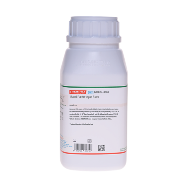
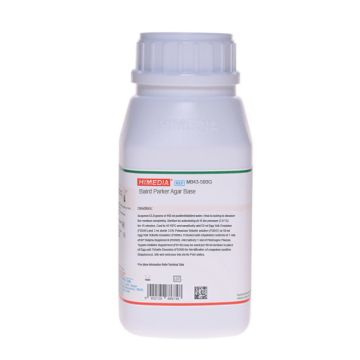
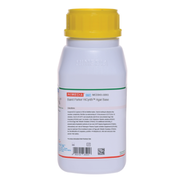
Baird Parker Agar Base
Baird Parker Agar Base with supplements is recommended for isolation and enumeration of coagulase positive Staphylococci from food and other materials. It is recommended by BIS under the specifications IS: 5887 (Part II) 1976.
Composition**
| Ingredients | Gms/Litre |
|---|---|
| Casein enzymic hydrolysate | 10.000 |
| Meat extract | 5.000 |
| Yeast extract | 1.000 |
| Glycine | 12.000 |
| Sodium pyruvate | 12.000 |
| Lithium chloride | 5.000 |
| Agar | 20.000 |
Final pH (at 25°C): 7.0±0.2
**Formula adjusted, standardized to suit performance parameters
Directions
Suspend 65 grams in 950 ml distilled water. Heat to boiling to dissolve the medium completely. Sterilize by autoclaving at 15 lbs pressure (121°C) for 15 minutes. Cool to 50°C and aseptically add 50 ml concentrated Egg Yolk Emulsion (FD045) and 3 ml sterile 3.5% Potassium Tellurite solution (FD047) or 50 ml Egg Yolk Tellurite Emulsion (FD046). Mix well and pour into sterile Petri plates.
Warning: Lithium chloride is harmful. Avoid bodily contact and inhalation of vapours. On contact with skin wash with plenty of water immediately.
Principle And Interpretation
Baird Parker Agar was developed by Baird-Parker (1,2) from the Tellurite - glycine formulation of Zebovitz et al (3) for isolation of Staphylococcus aureus from foods.
This medium was found to be less inhibitory to Staphylococcus aureus than other media, at the same time being more selective (4, 5, 6). Subsequently it was officially adapted by the AOAC and is also recommended in USP for use in Microbial limit test (7). ISO Committtee(10) has recommended this medium for isolation and enumeration of Staphylococci. Present formulation having slightly increased amount of pyruvate is recommended by BIS for isolation of Staphylococcus aureus (11). However identity of Staphylococcus aureus isolated on Baird-Parker Agar must be confirmed with a coagulase reaction. Smith and Baird-Parker (8) found that the addition of 50 mg/l Sulphamethazine in the medium, suppresses the growth and swarming of Proteus species
Sodium pyruvate protects injured cells and helps recovery. Lithium chloride and potassium tellurite inhibit most of the contaminating microflora except Staphylococcus aureus. Glycine, pyruvate enhances growth of Staphylococcus. With the addition of egg yolk, the medium becomes yellow, opaque. Proteolytic bacteria produce a clear zone around colony in egg yolk containing media. A clear zone and grey-black colonies on this medium are diagnostic for coagulase positive Staphylococci.
Upon further incubation, an opaque zone is developed around colonies which can be due to lipolytic activity.
Baird-Parker Agar Base can also be used to detect coagulase activity by adding plasma fibrinogen mixture in place of egg yolk emulsion. 375 mg bovine fibrinogen, 2.5 ml rabbit plasma, 2.5 mg trypsin inhibitor and 2.5 mg potassium tellurite dissolved in 10 ml sterile distilled water and added to 90 ml sterile molten medium kept at 45-50°C (9). Mix well and pour into plates. On this medium Staphylococcal coagulase positive colonies are white to grey-black surrounded by an opaque zone of coagulase activity, within 24-40 hours incubation at 35°C. Reduction in tellurite is necessary because of absence of egg yolk emulsion. This results in translucent agar and white to grey coloured colonies of Staphylococci. For quantitative results, select 20 - 200 colonies. Count Staphylococcus aureus like colonies and test them for coagulase reaction. Report Staphylococcus aureus per gram of food. Regardless of the negative reactions, consider all doubtful colonies as Staphylococcus aureus and carry out further tests. Colonies of some contaminating organisms may digest the coagulase halo reaction.
Quality Control
Appearance Cream to yellow coloured homogeneous free flowing powder
Gelling Firm, comparable with 2.0% agar gel.
Colour and Clarity of prepared medium Basal medium: Yellow coloured clear to slightly opalescent gel. After addition of Egg Yolk Emulsion and Tellurite Emulsion: Yellow coloured opaque gel forms in Petri plates.
Reaction Reaction of 6.5% w/v aqueous solution at 25°C. pH : 7.0±0.2
pH 6.80-7.20
Cultural Response Cultural characteristics observed with added Egg yolk emulsion and Tellurite Emulsion (FD045 and FD052), after an incubation at 35-37°C for 24-48 hours.
Cultural Response
| Organism | Inoculum (CFU) | Growth | Recovery | Colour of colony | Lecithinase |
|---|---|---|---|---|---|
| Bacillus subtilis ATCC 6633 | 50-100 | none - poor | <=10% | ||
| Micrococcus luteus ATCC 10240 | 50-100 | fair-good | 30-40% | shades of brown-black (very small) | Negative |
| Proteus mirabilis ATCC 25933 | 50-100 | good - luxuriant | >=50% | brown - black | Negative |
| Staphylococcus epidermidis ATCC 12228 | 50-100 | fair-good | 30-40% | black | Negative |
| Escherichia coli ATCC 25922 | 50-100 | none-poor | <=10% | ||
| Staphylococcus aureus ATCC 25923 | 50-100 | good - luxuriant | >=50% | grey-black shiny | Positive, opaque zone around the colony |
Storage and Shelf Life
Store below 30°C in tightly closed container and the prepared medium at 2 - 8°C. Use before expiry date on the label.
Reference
- Baird-Parker A.C., 1962, J. Appl. Bact., 25:12.
- Baird-Parker A.C. and Davenport E., 1965, J. Appl. Bact., 28:390.
- Zebovitz E., Evans J.B. and Niven C.F., 1955, J. Bact., 70:686.
- Tardio and Baer, 1971, J. Assoc. Off. Anal. Chem., 54:728.
- Baer, 1971, J. Assoc. Off. Anal. Chem., 54:732.
- J. Assoc. off. Anal. chem, 1971, 54:401.
- The United States Pharmacopoeia, 1995, 23rd rev., USP Convention, Rockville, Md.
- Smith B.A. and Baird-Parker A.C., 1964, J. Appl. Bact., 27:78.
- Beckers N. J. et al, 1984, Can. J. Microbiol., 30:470.
- International Organization for Standardization (ISO), 1983, Draft ISO/DIS 6888
- Bureau of Indian Standards IS: 5887 (Part II) 1976, (Second Reprint December 1994.
| Product Name | Baird Parker Agar Base |
|---|---|
| SKU | M043S |
| Product Type | Regular |
| Physical Form | Powder |
| Origin | Animal |
| Packaging type | HDPE |
| References | 1. Baird-Parker A. C., 1962, J. Appl. Bacteriol., 25:12. 2.Baird-Parker A. C. and Davenport E., 1965, J. Appl. Bacteriol., 28:390. 3.Zebovitz E., Evans J. B. and Niven C.F., 1955, J. Bacteriol., 70:686 . 4.Tardio and Baer, 1971, J. Assoc. Off. Anal. Chem., 54:728. 5.Baer, 1971, J. Assoc. Off. Anal. Chem., 54:732. 6.Assoc. off. Anal. Chem., 1971, 54:401. 7.Horwitz (Ed.), 2000, Official methods of analysis of AOAC International, 17th Ed., Vol. I., AOAC International,Gaithersburg, MD. 8.The United States Pharmacopoeia, 2018, The United States Pharmacopoeial Convention. Rockville, MD. 9.International Organization for Standardization (ISO), 1983, Draft ISO/DIS 6888. 10.Smith B. A. and Baird-Parker A.C., 1964, J. Appl. Bacteriol., 27:78. 11. Beckers N. J. et al, 1984, Can. J. Microbiol., 30:470.1 2.American Public Health Association, Standard Methods for the Examination of Dairy Products, 1978, 14th Ed., WashingtonD.C. 13.Salfinger Y., and Tortorello M.L. Fifth (Ed.), 2015, Compendium of Methods for the Microbiological Examination ofFoods, 5th Ed., American Public Health Association, Washington, D.C. 14..Wehr H. M. and Frank J. H., 2004, Standard Methods for the Microbiological Examination of Dairy Products, 17th Ed.,APHA Inc., Washington, D.C. 15.Isenberg, H.D. Clinical Microbiology Procedures Handb0ook. 2nd Edition. 16.Jorgensen,J.H., Pfaller , M.A., Carroll, K.C., Funke, G., Landry, M.L., Richter, S.S and Warnock., D.W. (2015)Manual of Clinical Microbiology, 11th Edition. Vol. 1. |
| Customized Product Available | No |


