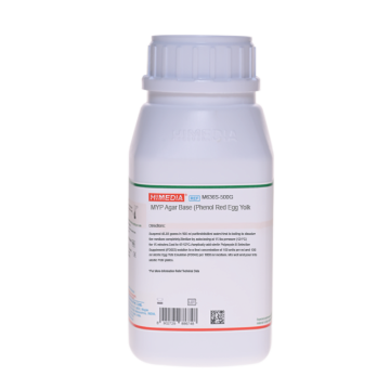
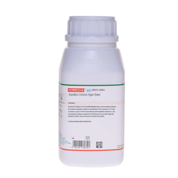
 Your enquiry has been submitted
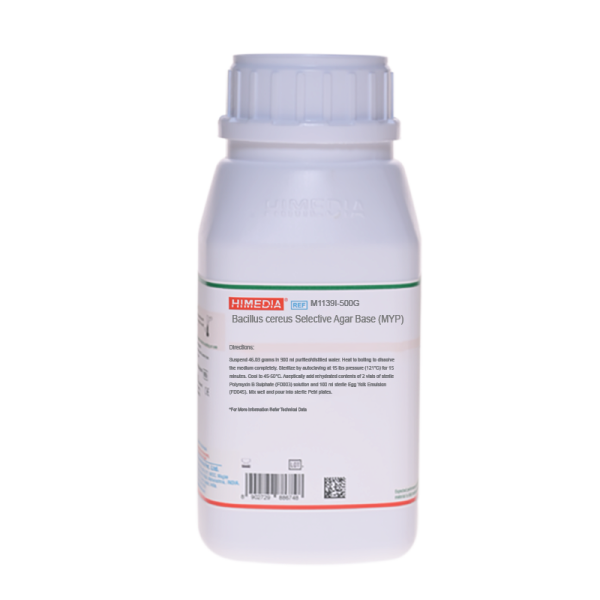
Bacillus cereus Selective Agar Base (MYP) ISO 7932
Intended Use:
Recommended for the isolation and identification of Bacillus species and pathogenic Staphylococci. The composition and performance criteria of this medium are as per the specification laid down in ISO 7932:2004/ Amd 1:2020, ISO 11133:2014 (E) & Amd:2020.
Composition**
ISO specifications: MYP
| Ingredients | g/L |
|---|---|
| Enzymatic digest of casein | 10.000 |
| Beef extract | 1.000 |
| D-Mannitol | 10.000 |
| Sodium chloride | 10.000 |
| Phenol red | 0.025 |
| Agar | 12.0-18.0 |
Polymyxin B sulphate: 50,000 units
Egg yolk emulsion: 100.00ml
Final pH (at 25°C): 7.2±0.2
Bacillus cereus Selective Agar Base (MYP) ISO 7932
| Ingredients | g/L |
|---|---|
| Tryptone | 10.000 |
| HM peptone B # | 1.000 |
| D-Mannitol | 10.000 |
| Sodium chloride | 10.000 |
| Phenol red | 0.025 |
| Agar | 15.000 |
FD003 - 2 vials
PolyB Selective Supplement: Polymyxin B sulphate 50,000 units
Egg Yolk Emulsion (FD045): Egg Yolk Emulsion 100 ml
Final pH (at 25°C): 7.2±0.2
**Formula adjusted, standardized to suit performance parameters
# Equivalent to Beef extract
Directions
Suspend 46.03 gram in 900 ml purified / distilled water. Heat to boiling to dissolve the medium completely. Sterilize by autoclaving at 15 lbs pressure (121°C) for 15 minutes. Cool to 45-50°C. Aseptically add rehydrated contents of 2 vials of sterile PolyB Selective Supplement (FD003) solution and 100 ml sterile Egg Yolk Emulsion (FD045). Mix well and pour into sterile Petri plates.
Principle And Interpretation
Bacillus cereus is ubiquitously present in soil, vegetation, water and dust. It has been isolated from a large variety of foods, pasteurized fresh milk and powdered milk (1-3) and processed foods. Under favourable conditions, the organism multiplies and causes gastrointestinal illness (4). It is implicated in two different forms of food poisoning; an emetic illness and a diarrhoeal illness. The emetic illness is mediated by a highly stable toxin that survives high temperature, exposure to trypsin, pepsin and pH extremes. The diarrhoeal illness is mediated by a heat and acid labile enterotoxin. Lecithinase activity is the key reaction in the differential identification of B.cereus, the most commonly encountered and important species in clinical laboratories, from the majority of the other Bacillus species. If unknown isolate produces lecithinase, B.cereus can be presumptively identified by also observing colonial morphology, hemolytic reactivity and motility tests. When present in large numbers in certain foodstuffs, B.cereus can produce metabolites responsible for the clinical symptoms of food poisoning (5). This medium differentiates B.cereus from other bacteria based on the basis of lecithinase activity, mannitol fermentation and resistance to polymyxin (FD003) (6,7). Bacillus cereus Selective Agar Base is recommended by the ISO committee for the enumeration of B.cereus (8,9). It contains tryptone and HM peptone B, which provide nitrogen source. Mannitol fermentation can be detected by phenol red, which yields yellow colour to the mannitol fermenting colonies due to acid production. Added egg yolk emulsion helps in differentiation of lecithinase producing colonies, which are surrounded by a zone of white precipitate. Addition of Polymyxin B Sulphate (FD003) helps to restrict growth of gram-negative bacteria such as Escherichia coli and Pseudomonas aeruginosa. These differentiating media allow differentiation of B.cereus from other Bacillus species by its inability to ferment mannitol and poor sporulation. B.cereus dissimilates egg yolk and gives rise to typical bacilli form colonies with reddish zones and white halos. Acid produced by organisms other than B.cereus often diffuse through the medium, making it difficult to distinguish between mannitol fermenters and non-fermenters. So it is advised to transfer the suspected colonies to a fresh medium to visualize the true reaction.
Type of specimen
Food and animal feeding stuffs
Specimen collection and handling
ISO 7932:2004
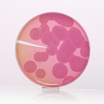
Prepare the test sample in accordance with the specific International Standard appropriate to the product concerned. Carefully spread the inoculum as quickly as possible over the surface of the agar plate without touching the sides of the dish with the spreader. Invert the prepared plates and incubate them for 18 h to 24 h in an incubator set at 30 °C. If colonies are not clearly visible, incubate the plates for an additional 24 h before counting. The presumptive colonies are large, pink, indicating that mannitol fermentation has not occurred and generally surrounded by a zone of precipitation indicating the production of lecithinase.
Confirmation: Streak, stab or spot the selected colonies onto the surface of sheep blood agar in a manner which allows good interpretation of the haemolysis reaction. Incubate at 30 °C for 24 h ± 2 h and interpret the haemolysis reaction.
Warning and Precautions:
Read the label before opening the container. Wear protective gloves/protective clothing/eye protection/ face protection. Follow good microbiological lab practices while handling specimens and culture. Standard precautions as per established guidelines should be followed while handling specimens. Safety guidelines may be referred in individual safety data sheets.
Limitations :
- Individual organisms differ in their growth requirement and may show variable growth patterns on the medium.
- Each lot of the medium has been tested for the organisms specified on the COA. It is recommended to users to validate the medium for any specific microorganism other than mentioned in the COA based on the user's unique requirement.It is advised to transfer the suspected colonies to a fresh medium to visualize the true reaction.
Performance and Evaluation
Performance of the medium is expected when used as per the direction on the label within the expiry period when stored at recommended temperature.
Quality Control
Appearance: Light yellow to pinkish purple homogeneous free flowing powder
Gelling: Firm, comparable with 1.5% Agar gel
Colour and Clarity of prepared medium: Basal medium: Red coloured clear to slightly opalescent gel. After Addition of Egg Yolk Emulsion (FD045): Light orange coloured opaque gel forms in Petri plates
Reaction: Reaction of 4.6% w/v aqueous solution at 25°C. pH: 7.2±0.2
pH: 7.0-7.40
Cultural Response
Productivity: Cultural characteristics observed with added Egg Yolk Emulsion (FD045) and PolyB Selective Supplement (FD003) after an incubation at 30 ± 1°C for 24 ± 3 to 44 ± 4 hours. Recovery rate is considered as 100% for bacteria growth on Reference Medium - Soyabean Casein Digest Agar.
Specificity: Cultural characteristics observed with added Egg Yolk Emulsion (FD045) and PolyB Selective Supplement (FD003) after an incubation at 30 ± 1°C for 24 ± 3 to 44 ± 4 hours.
Selectivity: Cultural characteristics observed with added Egg Yolk Emulsion (FD045) and PolyB Selective Supplement (FD003) after an incubation at 30 ± 1°C for 24 ± 3 to 44 ± 4 hours.
| Organism | Inoculum (CFU) | Growth | Recovery | Characteristic reaction |
|---|---|---|---|---|
| Productivity | ||||
| Bacillus cereus ATCC 11778 (00001*) | 50-100 | luxuriant | >=50% | Pink colonies with precipitation halo |
| Selectivity | ||||
| Escherichia coli ATCC 25922 (00013*) | >=104 | Inhibition | ||
| Specificity | ||||
| $ Bacillus spizizenii ATCC 6633 (00003*) | 103-104 | luxuriant | yellow colonies with precipitation halo | |
Key: *Corresponding WDCM numbers. $ - Formerly known as Bacillus subtilis subsp. spizizenii
Storage and Shelf Life
Store between 10-30°C in a tightly closed container and the prepared medium at 2-8°C. Use before expiry date on the label. On opening, product should be properly stored dry, after tightly capping the bottle in order to prevent lump formation due to the hygroscopic nature of the product. Improper storage of the product may lead to lump formation. Store in dry ventilated area protected from extremes of temperature and sources of ignition. Seal the container tightly after use.
Product performance is best if used within stated expiry period.
Disposal
User must ensure safe disposal by autoclaving and/or incineration of used or unusable preparations of this product. Follow established laboratory procedures in disposing of infectious materials and material that comes into contact with sample must be decontaminated and disposed of in accordance with current laboratory techniques (4,10).
Reference
- Bergdoll M. S., 1981, Clin. Microbiol. Newsletter 3: 85-87.
- Centers for Disease Control: Bacillus cereus- Maine, MMWR, 35: 408-410, 1986.
- Donovan K. O., 1958, J. Appl. Bacteriol., 21:100.
- Isenberg, H.D. Clinical Microbiology Procedures Handbook 2nd Edition.
- Mossel D. A. A., Koopman M. J. and Jongerium E., 1967, Appl. Microbiol, 15:650.
- Nygren B., 1962, Acta Path. Microbiol. Scand., 56: Suppl1.
- Salfinger Y., and Tortorello M.L., 2015, Compendium of Methods for the Microbiological Examination of Foods, 5th Ed., American Public Health Association, Washington, D.C.
- Microbiology of food and animal feeding stuffs — Horizontal method for the enumeration of presumptive Bacillus cereus — Colony-count technique at 30 degrees C — Amendment 1: Inclusion of optional tests. ISO 7932:2004/Amd 1:2020
- Microbiology of food, animal feeding stuffs and water— Preparation, production, storage and performance testing of culture media ISO 11133:2014 (E) & Amd. :2020
- Jorgensen, J.H., Pfaller, M.A., Carroll, K.C., Funke, G., Landry, M.L., Richter, S.S and Warnock., D.W. (2015) Manual of Clinical Microbiology, 11th Edition. Vol. 1.
| Product Name | Bacillus cereus Selective Agar Base (MYP) ISO 7932 |
|---|---|
| SKU | M1139I |
| Product Type | Regular |
| Physical Form | Powder |
| Origin | Animal |
| Packaging type | HDPE |
| References | 1.Bergdoll M. S., 1981, Clin. Microbiol. Newsletter 3: 85-87.2.Centers for Disease Control: Bacillus cereus- Maine, MMWR, 35: 408-410, 1986.3.Mossel D. A. A., Koopman M. J. and Jongerium E., 1967, Appl. Microbiol, 15:650.4.Downes F. P. and Ito K., (Eds.), 2001, Compendium of Methods for the Microbiological Examination of Foods, 4th Ed.,APHA, Washington, D.C.5.Nygren B., 1962, Acta Path. Microbiol. Scand., 56: Suppl. 1.6.Donovan K. O., 1958, J. Appl. Bacteriol., 21:100.7.Colimer A. R., 1948, J. Bacteriol., 55:777.8.Rhodehamel J. and Harmon S. M., 1995, FDA Bacteriological Analytical Manual, 8th Ed., AOAC International,Gaithersburg, Md.9.International Organization for Standardization (ISO), 1993, Draft ISO/DIS 7932. |
| Customized Product Available | No |


