 Your enquiry has been submitted
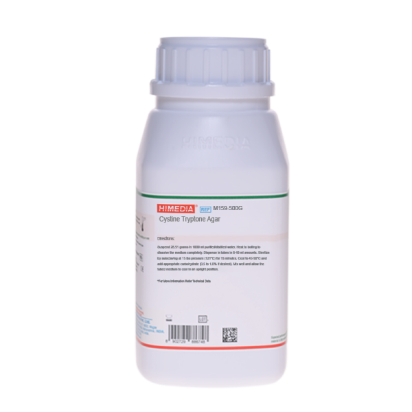
Cystine Tryptone Agar
Intended Use:
Recommended for maintenance, subculturing, detection of motility and fermentation studies with the addition of various carbohydrates.
Composition**
| Ingredients | Gms / Litre |
|---|---|
| Tryptone | 20.000 |
| L-Cystine | 0.500 |
| Sodium chloride | 5.000 |
| Sodium sulphite | 0.500 |
| Phenol red | 0.017 |
| Agar | 2.500 |
Final pH ( at 25°C) 7.3±0.2
**Formula adjusted, standardized to suit performance parameters
Directions
Suspend 28.51 grams in 1000 ml purified / distilled water. Heat to boiling to dissolve the medium completely. Dispense in tubes in 8-10 ml amounts. Sterilize by autoclaving at 15 lbs pressure (121°C) for 15 minutes. Cool to 45-50°C and add appropriate carbohydrate (0.5 to 1.0% if desired). Mix well and allow the tubed medium to cool in an upright position.
Principle And Interpretation
Cystine Tryptone Agar appropriates for the propagation and maintenance of bacteria even the fastidious ones without addition of additives. This formulation was developed by Vera as a simple semisolid medium for the identification and maintenance of the Gonococcus and other bacteria (14). In this medium by deep stab, a lot of cultures can be maintained including fastidious organisms like Brucella, Corynebacterium, Pasteurella, Pneumococcus and Streptococcus without added enrichments (1,11, 12) for longer periods when stored at appropriate temperatures. Even some light-sensitive anaerobic microorganisms can grow in this medium without special conditions though in reduced atmospheres, they give ideal growth. This medium has its maximum efficiency when freshly prepared, but it can be stored for long period of time, taking care to avoid dehydration. To achieve this, screw caps or proper sealing are strongly recommended. Anaerobic organisms like Actinomyces bovis, Bacteroides funduliformis and Leptotrichia (8) grow well in this medium in presence of CO2. With added carbohydrate, it may be used to study sugar fermentation of microorganisms that do not grow on phenol red classical media. Acidification can be easily observed with the change in colour of phenol red indicator. In semisolid agar, acid reactions are easily detected because the acid formed is not immediately diffused throughout the entire culture as in broth. Most cultures show an alkaline reaction when no fermentable carbohydrate is present. Motility can be readily detected in the semisolid medium (13). Motile cultures show growth away from the line of inoculation. Non-motile organisms grow in the inoculated area, along the stab line while the surrounding area remains clear.
Tryptone, L-cystine supplies the nutrients necessary to support the growth of fastidious microorganism. Carbohydrate fermentation is detected by a visible colour change of the medium due to the incorporation of the pH indicator dye, phenol red. When the organism metabolizes the carbohydrate present, organic acids are produced and the medium becomes acidified. However, the peptones present in the medium are also degraded by the bacteria present and yield substances that are alkaline in pH. The phenol red indicator changes from reddish-orange to yellow when the amount of acid produced by carbohydrate fermentation is greater than the alkaline end products of the peptone degradation. The colour change with phenol red occurs around pH 6.8, near the original pH of the medium.
Only the surface of the tubed medium is inoculated in case of fermentation studies of the genus Neisseria. For facultative organisms, such as Streptococci and strictly anaerobic organisms inoculation is done by stabbing the center of the medium with an inoculating needle to about ½ the depth of the medium. Incubate with loosened caps aerobically or anaerobically depending upon the organisms being tested. Neisseria should be incubated with loose caps (7); if incubated in CO2 incubator (3,10) or with tight caps in non-CO2 incubator (3). For more rapid growth and also for more rapid fermentation reactions, anaerobic cultures preferably should be incubated in the presence of CO2 as well as hydrogen or nitrogen.
Some strict anaerobes fail to grow or grow poorly in the absence of CO2. A yellow colour either in the upper one-third or throughout the medium indicates acid production due to carbohydrate fermentation. A red (alkaline) to orange (neutral) colour indicates that the carbohydrate has not been degraded and that only the peptone has been utilized. Inoculated medium (without carbohydrate) also exhibits a red to orange colour. This medium requires a heavy inoculum (9). Prolonged incubation may lead to changes in pH indicator or abnormal lactose/sucrose reactions with Neisseria pathogens (2,4). Neisseria species usually produce acid only in the area of stabs (upper third). If there is a strong acid (yellow color) throughout the medium, a contaminating organism may be present. Gram stain and oxidase test should be performed on the growth to confirm the presence of Neisseria species (9).
Type of specimen
Isolated Microorganism
Specimen Collection and Handling:
For Isolated Microorganism samples, follow appropriate techniques for sample collection, processing as per guidelines and local standards.(14) After use, contaminated materials must be sterilized by autoclaving before discarding.
Warning and Precautions :
Read the label before opening the container. Wear protective gloves/protective clothing/eye protection/ face protection. Follow good microbiological lab practices while handling specimens and culture. Standard precautions as per established guidelines should be followed while handling specimens. Safety guidelines may be referred in individual safety data sheets.
Limitations :
- Lack of sufficient inoculum may lead to false results.
- Do not inoculate to the bottom of the tube; improper inoculation may lead to weak acid reactions, thus creating difficulty in test interpretation.
Performance and Evaluation
Performance of the medium is expected when used as per the direction on the label within the expiry period when stored at recommended temperature.
Quality Control
Appearance Light yellow to light pink homogeneous free flowing powder
Gelling Semisolid, comparable with 0.25% Agar gel.
Colour and Clarity of prepared medium Red coloured, clear to slightly opalescent gel forms in tubes as butts
Reaction Reaction of 2.85% w/v aqueous solution at 25°C. pH : 7.3±0.2
pH 7.10-7.50
Cultural Response Cultural characteristics observed after an incubation at 35-37°C for 4-18 hours or longer if necessary.
| Organism | Inoculum (CFU) | Growth | Motility | Acid in presence of Dextrose |
|---|---|---|---|---|
| Escherichia coli ATCC 25922 (00013*) | 50-100 | good-luxuriant | positive, growth away from stabline causing turbidity | positive reaction, yellow colour |
| Neisseria gonorrhoeae ATCC 19424 | 50-100 | good | negative, growth along the stabline, surrounding medium remains clear | positive reaction, yellow colour |
| Neisseria meningitidis ATCC 13090 | 50-100 | good | negative, growth along the stabline, surrounding medium remains clear | positive reaction, yellow colour |
| Streptococcus pneumoniae ATCC 6303 | 50-100 | good | negative, growth along the stabline, surrounding medium remains clear | positive reaction, yellow colour |
Key : *Corresponding WDCM numbers.
Storage and Shelf Life
Store between 10-30°C in a tightly closed container and the prepared medium at 20-30°C. Use before expiry date on the label. On opening, product should be properly stored dry, after tightly capping the bottle in order to prevent lump formation due to the hygroscopic nature of the product. Improper storage of the product may lead to lump formation. Store in dry ventilated area protected from extremes of temperature and sources of ignition. Seal the container tightly after use. Product performance is best if used within stated expiry period.
Disposal
User must ensure safe disposal by autoclaving and/or incineration of used or unusable preparations of this product. Follow established laboratory procedures in disposing of infectious materials and material that comes into contact with clinical sample must be decontaminated and disposed of in accordance with current laboratory techniques (5,6).
Reference
- Alford, Wiese and Gunter, 1955, J. Bacteriol. 69:518.
- Applebaum and Lawrence, 1979, J. Clin. Microbiol. 9:598.
- Baron E. J., Peterson and Finegold S. M., Bailey & Scotts Diagnostic Microbiology, 9th Ed., 1994, Mosby-Year Book, Inc., St. Louis, Mo.
- Faur, Weisburd and Wilson, 1975, J. Clin. Microbiol. 1:294.
- Isenberg, H.D. Clinical Microbiology Procedures Handbook 2nd Edition.
- Jorgensen, J.H., Pfaller, M.A., Carroll, K.C., Funke, G., Landry, M.L., Richter, S.S and Warnock., D.W. (2015) Manual of Clinical Microbiology, 11th Edition. Vol. 1.
- Kellogg, 1974, Manual of Clinical Microbiology, 2nd Ed. American Society for Microbiology, Washington D.C.
- Kroeger and Sibal, 1961, J. Bacteriol. 50:581.
- MacFaddin J. F., 1985, Media for Isolation-Cultivation-Identification -Maintenance of Medical Bacteria, Vol. I, Williams and Wilkins, Baltimore.
- Morse and Knapp, 1987, 7th Ed., American Public Health Association,Washington D.C.
- Myers and Koshy, 1962, Am. J. Public Health, 96:75.
- Peterson and Hartsell, 1955, J. Inf. Dis., 96:75.
- Vera and Petran, 1954, Bull. Nat. Assoc. Clin. Labs., 5:90
- Vera H. D., 1948, J. Bacteriol. 55:531.
- Yu and Washington, 1985, Laboratory Procedures in Clinical Microbiology, 2nd Ed., Springer - Verlag, New York, N.Y.
| Product Name | Cystine Tryptone Agar |
|---|---|
| SKU | M159 |
| Product Type | Regular |
| Physical Form | Powder |
| Origin | Animal |
| Packaging type | HDPE |
| References | 1. Vera H. D., 1948, J. Bacteriol. 55:531. 2.Peterson and Hartsell, 1955, J. Inf. Dis., 96:75. 3.Myers and Koshy, 1962, Am. J. Public Health, 96:75. 4.Alford, Wiese and Gunter, 1955, J. Bacteriol. 69:518. 5.Kroeger and Sibal, 1961, J. Bacteriol. 50:581. 6.Vera and Petran, 1954, Bull. Nat. Assoc. Clin. Labs., 5:90 8.Faur, Weisburd and Wilson, 1975, J. Clin. Microbiol. 1:294. 9.Applebaum and Lawrence, 1979, J. Clin. Microbiol. 9:598. 10.Kellogg, 1974, Manual of Clinical Microbiology, 2nd Ed. American Society for Microbiology, Washington D.C. 11. Yu and Washington, 1985, Laboratory Procedures in Clinical Microbiology, 2nd Ed., Springer - Verlag, New York, N.Y.1 2.Morse and Knapp, 1987, 7th Ed., American Public Health Association,Washington D.C.1 3.Baron E. J., Peterson and Finegold S. M., Bailey & Scotts Diagnostic Microbiology, 9th Ed., 1994, Mosby-Year Book,Inc., St. Louis, Mo.1 4.Isenberg, H.D. Clinical Microbiology Procedures Handbook 2nd Edition. 15.Jorgensen,J.H., Pfaller, M.A., Carroll, K.C., Funke, G., Landry, M.L., Richter, S.S and Warnock., D.W. (2015)Manual of Clinical Microbiology, 11th Edition. Vol. 1. |
| Customized Product Available | No |


