 Your enquiry has been submitted
Something problem with the server. Try again.
Your enquiry has been submitted
Recommended for identification, differentiation and confirmation of enteric bacteria from specimens such as urine which may contain large number of Proteus species as well as potentially pathogenic gram-positive organisms.
| Ingredients | g/L |
|---|---|
| Peptone | 18.000 |
| Tryptone | 4.000 |
| HM Peptone B# | 6.000 |
| Chromogenic mixture | 12.440 |
| Agar | 15.000 |
Final pH (at 25°C): 7.2±0.2
**Formula adjusted, standardized to suit performance parameters
# -Equivalent to Beef extract
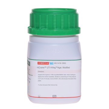
Suspend 55.44 gram in 1000 ml purified/distilled water. Heat to boiling to dissolve the medium completely. Sterilize by autoclaving at 15 lbs pressure (121°C) for 15 minutes. Cool to 45-50°C. Mix well and pour into sterile Petri plates.
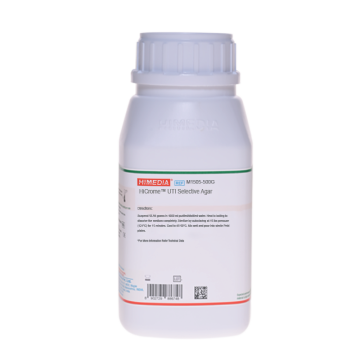
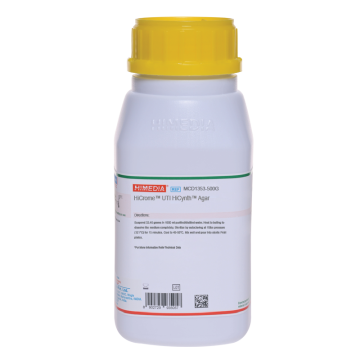
HiCrome® UTI Agar, Modified is formulated on the basis of work carried out by Pezzlo (1), Wilkie et al (2), Friedman et al (3), Murray et al (4), Soriano and Ponte (5) and Merlino et al (6). These media is the modification of HiCrome® UTI Agar (M1353), which can be used in place of MacConkey Agar for isolation and confirmation of various microorganisms. It facilitates and expedites the identification of some gram-negative bacteria and some gram-positive bacteria on the basis of different contrasted colony colours produced by reactions of genus or species specific enzymes with two chromogenic substrates.
Enzymes produced by Enterococcus species, Escherichia coli and coliforms cleave the chromogenic substrates incorporated in the medium. Presence of rich source of phenylalanine and tryptophan from peptone and tryptone provides an indication of tryptophan deaminase activity, revealed with TDA Reagent (R036) indicating the presence of Proteus species, Morganella species and Providencia species, which appear brown. One chromogenic substrate is cleaved by β-glucosidase possessed by Enterococci resulting in formation of blue colonies. E.coli produce purple-magenta colonies due to the enzyme β-D-galactosidase which cleaves the other chromogenic substrate. Further confirmation of E.coli can be done by performing indole test using DMACA Reagent (R035). Also, some strains of Enterobacter cloacae lacking β-glucosidase show pink-colonies indistinguishable from E.coli. The DMACA reagent for indole test (should be performed on filter paper) distinguishes between E.coli and Enterobacter, and also between Proteus mirabilis and other species. Coliforms produce purple coloured colonies due to cleavage of both the chromogenic substrates Peptone, HM Peptone B and tryptone provides nitrogenous, carbonaceous compounds and other essential growth nutrients.
Clinical samples : urine, faeces, etc.; Food samples; Water samples.
For clinical samples follow appropriate techniques for handling specimens as per established guidelines (7,8).
For food and dairy samples, follow appropriate techniques for sample collection and processing as per guidelines (9,10). For water samples, follow appropriate techniques for sample collection, processing as per guidelines and local standards (11).
After use, contaminated materials must be sterilized by autoclaving before discarding.
In Vitro diagnostic use. For professional use only. Read the label before opening the container. Wear protective gloves/protective clothing/eye protection/face protection. Follow good microbiological lab practices while handling specimens and culture. Standard precautions as per established guidelines should be followed while handling clinical specimens. Safety guidelines may be referred in individual safety data sheets.
Performance of the medium is expected when used as per the direction on the label within the expiry period when stored at recommended temperature.
Appearance
Cream to yellow homogeneous free flowing powder
Gelling
Firm, comparable with 1.5% Agar gel
Colour and Clarity of prepared medium
Light amber coloured, clear to slightly opalescent gel forms in Petri plates
Reaction
Reaction of 5.54% w/v aqueous solution at 25°C. pH : 7.2±0.2
pH
7.00-7.40
Cultural characteristics observed after an incubation at 35-37°C for 24 hours.
| Organism | Inoculum (CFU) | Growth | Recovery | Colour of Colony | TDA (add 1-2 drops of TDA reagent) | DMACA (transfer colony on filter paper dipped in DMACA Reagent) |
|---|---|---|---|---|---|---|
| Escherichia coli ATCC 25922 (00013*) | 50-100 | luxuriant | >=70% | Purple to magenta | negative reaction | positive reaction, formation of blue purple colour around growth |
| Enterococcus faecalis ATCC 29212 (00087*) | 50-100 | luxuriant | >=70% | blue-green (small) | negative reaction | negative reaction |
| Klebsiella pneumoniae ATCC 13883 (00097*) | 50-100 | luxuriant | >=70% | blue to purple, mucoid | negative reaction | negative reaction |
| Proteus mirabilis ATCC 12453 | 50-100 | luxuriant | >=70% | light brown | positive reaction, development of brown colouration | negative reaction |
| Pseudomonas aeruginosa ATCC 27853 (00025*) | 50-100 | luxuriant | >=70% | colourless (greenish pigment may be observed) | negative reaction | negative reaction |
| Staphylococcus aureus subsp. aureus ATCC 25923 (00034*) | 50-100 | luxuriant | >=70% | golden yellow | negative reaction | negative reaction |
Key : (*) Corresponding WDCM numbers.
Store between 15-25°C in a tightly closed container and the prepared medium at 2-8°C. Use before expiry date on the label. On opening, product should be properly stored dry, after tightly capping the bottle in order to prevent lump formation due to the hygroscopic nature of the product. Improper storage of the product may lead to lump formation. Store in dry ventilated area protected from extremes of temperature and sources of ignition Seal the container tightly after use. Product performance is best if used within stated expiry period.
User must ensure safe disposal by autoclaving and/or incineration of used or unusable preparations of this product. Follow established laboratory procedures in disposing of infectious materials and material that comes into contact with clinical sample must be decontaminated and disposed of in accordance with current laboratory techniques (7,8).
| Product Name | HiCrome® UTI Agar, Modified |
|---|---|
| SKU | M1418 |
| Product Type | HiCrome™ |
| Physical Form | Powder |
| Origin | Animal |
| Packaging type | HDPE |
| References | 1.Baird R.B., Eaton A.D., and Rice E.W., (Eds.), 2015, Standard Methods for the Examination of Water and Wastewater, 23rd ed., APHA, Washington, D.C. 2.Friedman M.P. et al. (1991), Journal of Clinical Microbiology, 29:2385-238 |