 Your enquiry has been submitted
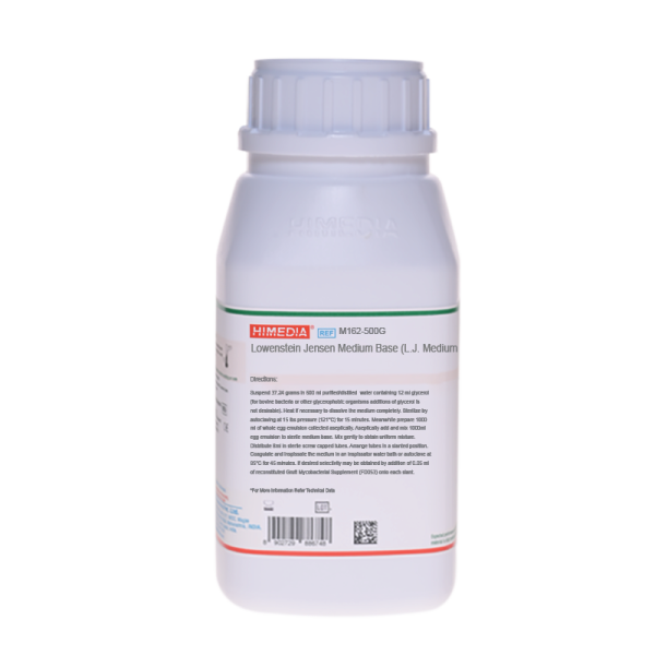
Lowenstein Jensen Medium Base (L.J. Medium)
Intended Use
Recommended for isolation and cultivation of Mycobacterium species.
Composition**
| Ingredients | g / 600 ml |
|---|---|
| L-Asparagine | 3.600 |
| Potassium dihydrogen phosphate | 2.400 |
| Magnesium sulphate | 0.240 |
| Magnesium citrate | 0.600 |
| Potato starch, soluble | 30.000 |
| Malachite green | 0.400 |
**Formula adjusted, standardized to suit performance parameters
Directions
Suspend 37.24 grams in 600 ml purified / distilled water containing 12 ml glycerol (for bovine bacteria or other glycerophobic organisms additions of glycerol is not desirable). Heat if necessary to dissolve the medium completely. Sterilize by autoclaving at 15 lbs pressure (121°C) for 15 minutes. Cool to 45-50°C. Meanwhile prepare 1000 ml of whole egg emulsion collected aseptically. Aseptically add and mix egg emulsion base and Gruft Selective Supplement (FD053) (if desired) gently to obtain uniform mixture. Distribute in sterile screw capped tubes. Arrange tubes in a slanted position. Coagulate and inspissate the medium in an inspissator water bath or autoclave at 85°C for 45 minutes.
Principle And Interpretation
Solid media used for isolation and cultivation of Mycobacteria are either egg-based or agar-based. Egg-based media contain whole eggs or egg yolk, potato flour, salts and glycerol and are solidified by inspissation. Of the egg-based media, LowensteinJensen Medium is most commonly used (1). L.J. Medium was originally formulated by Lowenstein, containing congo red and malachite green dyes (2). Jensen (3) modified Lowensteins medium by altering the citrate and phosphate contents, eliminating the congo red dye and by increasing the malachite green concentration. Gruft (4,5) further modified L. J. Medium with the addition of two antimicrobics to increase selectivity. This medium supports the growth of a wide variety of Mycobacteria and can also be used for niacin testing (6).
Penicillin and Nalidixic acid (FD053) along with malachite green prevents growth of the majority of contaminants surviving decontamination of the specimen while encouraging earliest possible growth of Mycobacteria. RNA (FD053) acts as stimulant and help to increase the isolation rate of Mycobacteria. Do not add glycerol to the medium if bovine or other glycerophobic strains are to be cultured (7). Malachite green serves as an inhibitor and also as pH indicator. Formation of blue zone indicates a decrease in pH by gram-positive contaminants (e.g. Streptococci) and yellow zones of dye destruction by gram-negative bacilli. Proteolytic contaminants cause localized or complete digestion of medium. Hardy et al (8) recommended each specimen to be inoculated and incubated in triplicate,
- To identify saprophytes at room temperature (25°C).
- To identify presence or absence of pigmentation by photochromogenes and scotochromogenes at 35°C alternately in light and dark as per the type of organism.
Routinely cultivation is carried out aerobically at 35°C.
Type of specimen
Clinical samples : Sputum
Specimen Collection and Handling
For clinical samples:Refer appropriate references for standard test procedures of decontamination and isolation (1,9-12). After use, contaminated materials must be sterilized by autoclaving before discarding.
Warning and Precautions
In Vitro diagnostic use only. For professional use only. Read the label before opening the container. Wear protective gloves/protective clothing/eye protection/face protection. Follow good microbiological lab practices while handling specimens and culture. Standard precautions as per established guidelines should be followed while handling clinical specimens. Safety guidelines may be referred in individual safety data sheets.
Limitations
- This medium is general purpose medium and may not support the growth of fastidious organisms.
- Certain gram-positive contaminants (e.g. Streptococci ) and gram-negative bacilli may grow on the medium.
- Certain Saprophytes may also grow on the medium .
- Proteolytic contaminants cause localized or complete digestion of medium.
Performance and Evaluation
Performance of the medium is expected when used as per the direction on the label within the expiry period when stored at recommended temperature.
Quality Control
Appearance Greenish blue to peacock blue homogeneous free flowing powder
Colour and Clarity of prepared medium The mixture of sterile basal medium and whole egg emulsion, when inspissated, coagulates to yield pale bluish green coloured, opaque smooth slants.
Cultural Response Cultural characteristics observed in presence of 5-10% CO2, with added egg emulsion base, after an incubation at 35-37ºC for 2-4 weeks.
| Organism | Growth | Growth with Gruft Selective Supplement (FD053) | Colony Characteristic |
|---|---|---|---|
| Mycobacterium avium ATCC 25291 | luxuriant | good-luxuriant | smooth, non-pigmented colonies |
| Mycobacterium gordonae ATCC 14470 | luxuriant | good-luxuriant | smooth, yellow, orange colonies |
| Mycobacterium kansasii ATCC 12478 | luxuriant | good-luxuriant | photochromogenic, smooth to rough |
| Mycobacterium smegmatis ATCC 14468 | luxuriant | good-luxuriant | wrinkled,creamy white colonies |
| M. tuberculosis H37RV ATCC 25618 | luxuriant | good-luxuriant | granular, rough, warty, dry friable colonies |
Storage and Shelf Life
Store below 10-30°C in a tightly closed container and the prepared medium at 2-8°C. Use before expiry date on the label. On opening, product should be properly stored dry, after tightly capping the bottle in-order to prevent lump formation due to the hygroscopic nature of the product. Improper storage of the product may lead to lump formation. Store in dry ventilated area protected from extremes of temperature and sources of ignition Seal the container tightly after use. Product performance is best if used within stated expiry period.
Disposal
User must ensure safe disposal by autoclaving and/or incineration of used or unusable preparations of this product. Follow established laboratory procedures in disposing of infectious materials and material that comes into contact with clinical sample must be decontaminated and disposed of in accordance with current laboratory techniques (9-12).
Reference
- Murray P. R., Baron E. J., Jorgensen J. H., Pfaller M. A., Yolken R. H., (Eds.), 8th Ed., 2003, Manual of Clinical Microbiology, ASM, Washington, D.C.
- Lowenstein E., 1931, Zentralbl. Bakteriol. Parasitenkd. Infektionskr. Hyg. Abt. 1 Orig., 120:127.
- Jensen K. A., 1932, Zentralb. Bakteriol. Parasitenkd. Infektionskr. Hyg. Abt. I Orig., 125:222.
- Gruft, 1971, Health Lab. Sci., 8:79.
- Gruft, 1963, Am. Rev. Respir. Dis., 88:412.
- Boisvert H., 1960, Ann. Inst. Pasteur, 99:600.
- MacFaddin J. F., 1985, Media for Isolation-Cultivation-Identification-Maintenance of Medical Bacteria, Vol. 1, Williams and Wilkins, Baltimore.
- A.V. Hardy, et al, Am. J. Publ. Hlth. 48(1), 754 (1958)
- Isenberg, (Ed.), Clinical Microbiology Procedures Handbook 2nd Edition
- Jorgensen, J.H., Pfaller, M.A., Carroll, K.C., Funke, G., Landry, M.L., Richter, S.S and Warnock., D.W. (2015) Manual of Clinical Microbiology, 11th Edition. Vol. 1.
- Cernoch P., Enns R., Saubolle M. and Wallace R., 1994, Cumitech, 16A, Laboratory Diagnosis of the Mycobacterioses coord , Ed., Weissfeld , ASM, Washington, D. C.
- Forbes B. A., Sahm A. S. and Weissfeld D. F., Bailey & Scotts Diagnostic Microbiology, 10th Ed., 1998, Mosby, Inc., St. Louis, Mo.
| Product Name | Lowenstein Jensen Medium Base (L.J. Medium) |
|---|---|
| SKU | M162 |
| Product Type | Regular |
| Physical Form | Powder |
| Origin | Animal Free (Veg) |
| Packaging type | HDPE |
| References | 1.A.V. Hardy, et al, Am. J. Publ. Hlth. 48(1), 754 (1958)2.Boisvert H., 1960, Ann. Inst. Pasteur, 99:600. 3.Cernoch P., Enns R., Saubolle M. and Wallace R., 1994, Cumitech, 16A, Laboratory Diagnosis of the Mycobacterioses coord , Ed., Weissfeld , ASM, Washington, D. C. 4.Forbes B. A., Sahm A. S. and Weissfeld D. F., Bailey & Scotts Diagnostic Microbiology, 10th Ed., 1998, Mosby, Inc., St.Louis, Mo. 5.Gruft, 1971, Health Lab. Sci., 8:79. 6.Gruft, 1963, Am. Rev. Respir. Dis., 88:412. 7.Isenberg, (Ed.), Clinical Microbiology Procedures Handbook 2nd Edition 8.Jensen K. A., 1932, Zentralb. Bakteriol. Parasitenkd. Infektionskr. Hyg. Abt. I Orig., 125:222 9.Jorgensen,J.H., Pfaller, M.A., Carroll, K.C., Funke, G., Landry, M.L.,Richter, S.S and Warnock., D.W. (2015) Manual of Clinical Microbiology, 11th Edition. Vol. 1. 10.Lowenstein E., 1931, Zentralbl. Bakteriol. Parasitenkd. Infektionskr. Hyg. Abt. 1 Orig., 120:127. 11.MacFaddin J. F., 1985, Media for Isolation-Cultivation-Identification-Maintenance of Medical Bacteria, Vol. 1,Williams and Wilkins, Baltimore. 12.Murray P. R., Baron E. J., Jorgensen J. H., Pfaller M. A., Yolken R. H., (Eds.), 8th Ed., 2003, Manual of Clinical Microbiology, ASM, Washington, D.C. |
| Customized Product Available | No |


