 Your enquiry has been submitted
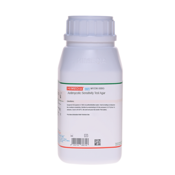
Antimycotic Sensitivity Test Agar
Intended Use:
Recommended for testing antimycotic sensitivity by diffusion method using antimycotic sensitivity discs.
Composition**
| Ingredients | Gms/Litre |
|---|---|
| Tryptone | 19.000 |
| Yeast extract | 10.000 |
| Dextrose (Glucose) | 20.000 |
| Sodium citrate | 10.000 |
| Disodium hydrogen phosphate | 1.000 |
| Agar | 25.000 |
Final pH (at 25°C): 6.6±0.2
**Formula adjusted, standardized to suit performance parameters
Directions
Suspend 85.0 grams in 1000 ml purified / distilled water. Heat to boiling to dissolve the medium completely. Sterilize by autoclaving at 15 lbs pressure (121°C) for 15 minutes. Cool to 45-50°C. Mix well and pour into sterile Petri plates.
Principle And Interpretation
Antimycotic Sensitivity Test Agar is recommended for testing the antimycotic activity by disc diffusion method (Sensitivity disc or MIC testing). The Clinical and Laboratory Standards Institute (CLSI) subcommittee on Antifungal Susceptibility Testing (6) has established interpretative break point for three drugs and Candida spp. The M44-A document (approved standard) became available in 2004. (5,10) Espinel-Ingroff (2)suggested that easier test modification to CLSI methods are desirable. Several workers have used agar diffusion method as an alternative approch to the CLSI methods. Stiller et al (9) observed a good correlation between MICs and growth inhibition zones for 5-FC. Pfaller et al (7) found a fluconazole disk test to be comparable to the MIC test. Barry and Brown (1) demonstrated good correlation between fluconazole disk test and MIC determined by either broth dilution or E Test. Espinel et al (2) and Pfaller et al (7) evaluated E Test and suggested that it correlates well with CLSI reference methods. However this agreement was species and medium dependent and they suggested the need for further optimisation of medium formulation. The medium contains tryptone and yeast extract which provide all essential growth nutrients like amino acids, vitamins, trace elements etc. Glucose serves as energy source. Disodium phosphate buffers the medium well.
Type of specimen
Isolated Microorganism
Specimen Collection and Handling
Technique:
Preparation of Inoculum:
- Inoculum is prepared by picking five distinct colonies of approximately 1mm from 24 hours old culture grown on Sabouraud Dextrose Agar (M063) and incubated at 35 ± 2°C. Colonies are suspended in 5ml of sterile 0.85% Saline.
- Vortex the resulting suspension and adjust the turbidity to yield 1 x 10⁶ - 5 x 10⁶ cells /ml (i.e. 0.5 McFarland standard).
Test Procedure:
- Prepare plates with Antimycotic sensitivity Test Agar for carrying out susceptibility of antifungal discs. The medium in the plates should be sterile and have a depth of about 4 mm.
- Dip a sterile non-toxic cotton swab on a wooden applicator into the standardized inoculum (turbidity so adjusted, as to obtain semi confluent growth on the petri plate) and rotate the soaked swab firmly against the upper inside wall of the tube to express excess fluid. Streak the entire agar surface of the plate with the swab three times, turning the plate at 60° angle between each streaking. Allow the inoculum to dry for 5 - 15 minutes with lid in place.
- Apply the discs using aseptic technique. Deposit the discs with centers at least 24 mm apart.
- Invert the plates and place in an incubator set to 35 ± 2°C within 15 minutes after the discs are applied.
- Examine each plate after 20 - 24 hours of incubation. If plate was satisfactorily streaked the resulting zones of inhibition will be uniformly circular and there will be a semi-confluent lawn of growth. Read at 48 hours only when insufficient growth is observed after 24 hours incubation.
Warning and Precautions
Read the label before opening the container. Wear protective gloves/protective clothing/eye protection/face protection. Follow good microbiological lab practices while handling specimens and culture. Standard precautions as per established guidelines should be followed while handling specimens. Safety guidelines may be referred in individual safety data sheets.
Limitations
- Freshly prepared medium plates must be used or it may result in erroneous results.
Performance and Evaluation
Performance of the medium is expected when used as per the direction on the label within the expiry period when stored at recommended temperature.
Quality Control
Appearance: Cream to yellow homogeneous free flowing powder
Gelling: Firm, comparable with 2.5% Agar gel.
Colour and Clarity of prepared medium: Amber coloured clear to slightly opalescent gel forms in Petri plates
Reaction: Reaction of 8.5% w/v aqueous solution at 25°C. pH : 6.6±0.2
pH: 6.40-6.80
Cultural Response
Average diameter of zone of inhibition observed after an incubation at 25-30°C for 48-72 hours.
| Organism | Growth | Zone of inhibition (Amphotericin B 50mcg) | Zone of inhibition (Nystatin 50mcg) | Zone of inhibition (Miconazole 50mcg) | Zone of inhibition (Miconazole 30mcg) | Zone of inhibition (Ketoconazole 50mcg) |
|---|---|---|---|---|---|---|
| Candida albicans ATCC 90028 | Luxuriant | 12-15mm | 19-23mm | 26-32mm | 22-26mm | 30-37mm |
| Candida parapsillosis ATCC 22019 | 11-18mm | 13-17mm | 19-23mm | 23-29mm | 13-17mm | 27-33mm |
Storage and Shelf Life
Store between 10-30°C in a tightly closed container and use freshly prepared medium. Use before expiry date on the label. On opening, product should be properly stored dry, after tightly capping the bottle in order to prevent lump formation due to the hygroscopic nature of the product. Improper storage of the product may lead to lump formation. Store in dry ventilated area protected from extremes of temperature and sources of ignition. Seal the container tightly after use. Product performance is best if used within stated expiry period.
Disposal
User must ensure safe disposal by autoclaving and/or incineration of used or unusable preparations of this product. Follow established laboratory procedures in disposing of infectious materials and material that comes into contact with sample must be decontaminated and disposed of in accordance with current laboratory techniques (3,4).
Reference
- Barry A.L. and S.D. Brown. 1996. J. Clin. Microbiol.34:2154-2157
- Espinel -Ingroff et al (1992) J.Clin. Microbiol. 30: 3138-3145
- Isenberg, H.D. Clinical Microbiology Procedures Handbook 2nd Edition
- Jorgensen, J.H. Pfaller, M.A., Carroll, K.C., Funke, G., Landry, M.L., Richter, S.S and Warnock., D.W. (2015) Manual Clinical Microbiology, 11th Edition. Vol. 1.
- Method for Antifungal Disk Diffusion Susceptibility Testing of Yeasts; Approved Guidelines-Second edition Vol.29 No.17, August-2009 CLSI document M44-A2. For more details refer to this volume
- National Committee for Clinical Laboratory Standards. 1997. Reference method for broth dilution antifungal susceptibility testing of yeasts; Approved standard M27-A.
- Pfaller M.A et al (1988) J.Clin. Microbiol 26: 1437-1441
- Rex, J.H. et al 1997 for the Subcommittee on Antifungal Susceptibility Testing of the National Committee for Clinical Laboratory Standards. Clin. Infect. Dis. 24: 235-247
- Stiller R.L. et al (1983) J.Infec. Dis 147: 1070-1076
- Zone Diameter Interpretive Standards, Corresponding Minimal Inhibitory Concentration (MIC) Interpretive Breakpoints, and Quality Control Limits for Antifungal Disk Diffusion Susceptibility Testing Of Yeasts, Third International Supplement CLSI document - M44-S3. National Committee for Clinical Laboratory Standards, villanova, Pa.
| Product Name | Antimycotic Sensitivity Test Agar |
|---|---|
| SKU | M1336 |
| Product Type | Regular |
| Physical Form | Powder |
| Origin | Animal |
| Packaging type | HDPE |
| References | 1.Rex, J.H. et al 1997 for the Subcommittee on Antifungal Susceptibility Testing of the National Committee for ClinicalLaboratory Standards. Clin. Infect. Dis. 24 : 235-2472.National Committee for Clinical Laboratory Standards. 199 |
| Customized Product Available | No |


