 Your enquiry has been submitted
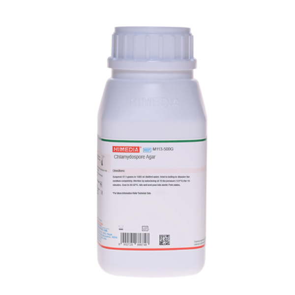
Chlamydospore Agar
Fungi -Yeasts and Molds#CC293D
Intended Use
Recommended for differentiation of Candida albicans from other Candida species on the basis of chlamydospore formation.
Composition**
| Ingredients | Gms / Litre |
|---|---|
| Ammonium sulphate | 1.000 |
| Monopotassium phosphate | 1.000 |
| Biotin | 0.000005 |
| Trypan blue | 0.100 |
| Purified polysaccharide | 20.000 |
| Agar | 15.000 |
| Final pH (at 25°C) | 5.1±0.2 |
**Formula adjusted, standardized to suit performance parameters
Directions
Suspend 37.1 grams in 1000 ml purified / distilled water. Heat to boiling to dissolve the medium completely. Sterilize by autoclaving at 15 lbs pressure (121°C) for 15 minutes. Cool to 45-50°C. Mix well and pour into sterile Petri plates.
Principle And Interpretation
Candida albicans is a diploid sexual fungus (a form of yeast), and the causitive agent of opportunistic oral and vaginal infections in humans (5). C. albicans is a commensal of skin, gastrointestinal and genitourinary tract. However, under certain conditions overgrowth of this results into oesopharyngeal candidiasis, vulvovaginal candidiasis and candidemia. Chlamydospores formation is the most differential characteristic of C. albicans (5). Chlamydospore Agar was specially designed for the differentiation of C.albicans from other species on the basis of chlamydospores formation. It is prepared according to the formula of Nickerson and Mankowshi (4).
Ammonium sulphate acts as sources of ions that simulate metabolism. Monopotassium phosphate provides buffering to the medium. Biotin provides the necessary vitamins required for metabolism. Purified polysaccharide acts as a source of carbon. Trypan blue is a vital dye absorbed selectively by the chlamydospores and imparts blue colour to chlamydospores, whereas the filaments are colourless.
Test for chlamydospores: Scratch cut mark like X onto the agar surface with inoculum using sterile needle. Aseptically place an alcohol-flamed and cooled cover slip onto the agar surface over the intersecting lines of the cut marks of X. Incubate plates at 20-25°C for 2-6 days. Temperature should not be higher than 25°C since it will not permit chlamydospore formation. Observe the plates under low power of microscope. After incubation, most strains of C.albicans and C.stellatoide will form typical chlamydospores. Chlamydospores will be seen along the edge of the cover slip. Chlamydospores are round, thick walled, blue coloured and at the terminal ends of hyphae.
Some C.albicans strains may lose their ability to produce chlamydospores after repeated subculturing.
Type of specimen
Food samples; Water samples
Specimen Collection and Handling
For food samples, follow appropriate techniques for sample collection and processing as per guidelines (1).
For water samples, follow appropriate techniques for sample collection, processing as per guidelines and local standards.(6)
After use, contaminated materials must be sterilized by autoclaving before discarding.
Warning and Precautions :
Read the label before opening the container. Wear protective gloves/protective clothing/eye protection/ face protection. Follow good microbiological lab practices while handling specimens and culture. Standard precautions as per established guidelines should be followed while handling specimens. Safety guidelines may be referred in individual safety data sheets.
Limitations :
- A temperature of 25°C is recommended for the foremost results.
Performance and Evaluation
Performance of the medium is expected when used as per the direction on the label within the expiry period when stored at recommended temperature.
Quality Control
Appearance Cream to blue homogeneous free flowing powder
Gelling Firm, comparable with 1.5% Agar gel
Colour and Clarity of prepared medium Blue coloured opaque gel forms in Petri plates
Reaction Reaction of 3.71% w/v aqueous solution at 25°C. pH: 5.1±0.2
pH 4.90-5.30
Cultural Response Cultural characteristics observed after an incubation at 20-25°C for 2-6 days.
| Organism | Growth | Chlamydospores |
|---|---|---|
| Candida albicans ATCC 10231 (00054*) | good-luxuriant | positive |
| Candida kruisei ATCC 24408 | good-luxuriant | negative |
| Candida minosa | good-luxuriant | negative |
| Candida tropicalis ATCC 1369 | good-luxuriant | negative |
Key: *Corresponding WDCM numbers.
Storage and Shelf Life
Store between 10-30°C in a tightly closed container and the prepared medium at 20-30°C. Use before expiry date on the label. On opening, product should be properly stored dry, after tightly capping the bottle in order to prevent lump formation due to the hygroscopic nature of the product. Improper storage of the product may lead to lump formation. Store in dry ventilated area protected from extremes of temperature and sources of ignition Seal the container tightly after use. Use before expiry date on the label.
Product performance is best if used within stated expiry period.
Disposal
User must ensure safe disposal by autoclaving and/or incineration of used or unusable preparations of this product. Follow established laboratory procedures in disposing of infectious materials and material that comes into contact with sample must be decontaminated and disposed of in accordance with current laboratory techniques (2,3).
Reference
- Baird R.B., Eaton A.D., and Rice E.W., (Eds.), 2015, Standard Methods for the Examination of Water and Wastewater, 23rd ed., APHA, Washington, D.C.
- Isenberg, H.D. Clinical Microbiology Procedures Handbook 2nd Edition.
- Jorgensen, J.H., Pfaller, M.A., Carroll, K.C., Funke, G., Landry, M.L., Richter, S.S and Warnock., D.W. (2015) Manual of Clinical Microbiology, 11th Edition. Vol. 1.
- Nickerson, 1953, J. Infect. Dis., 92:20
- Ryan K. J., Ray C. G., (Eds.), 2004, Sherris Medical Microbiology, 4th Ed., McGraw Hill.
- Salfinger Y., and Tortorello M.L., 2015, Compendium of Methods for the Microbiological Examination of Foods, 5th Ed., American Public Health Association, Washington, D.C.
| Product Name | Chlamydospore Agar |
|---|---|
| SKU | M113 |
| Product Type | Regular |
| Physical Form | Powder |
| Origin | Chemically defined (HiCynth™) |
| Packaging type | HDPE |
| References | 1. Baird R.B., Eaton A.D., and Rice E.W., (Eds.), 2015, Standard Methods for the Examination of Water andWastewater, 23rd ed., APHA, Washington, D.C. 2.Isenberg, H.D. Clinical Microbiology Procedures Handbook 2nd Edition. 3.Jorgensen, J.H., Pfaller, M.A., Carroll, K.C., Funke, G., Landry, M.L., Richter, S.S and Warnock., D.W.(2015) Manual of Clinical Microbiology, 11th Edition. Vol. 1.1 4.Nickerson, 1953, J. Infect. Dis., 92:20 5.Ryan K. J., Ray C. G., (Eds.), 2004, Sherris Medical Microbiology, 4th Ed., McGraw Hill. 6.Salfinger Y., and Tortorello M.L., 2015, Compendium of Methods for the Microbiological Examination of Foods, 5thEd., American Public Health Association, Washington, D.C. |
| Customized Product Available | No |


