 Your enquiry has been submitted
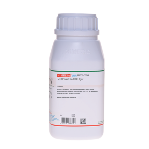
MUG Violet Red Bile Agar
Lactose fermenters#CC293D
Intended Use
Recommended for the detection and enumeration of coliform organisms by a fluorogenic method.
Composition
| Ingredients | Gms / Litre |
|---|---|
| Peptone | 7.000 |
| Yeast extract | 3.000 |
| Bile salts mixture | 1.500 |
| Lactose | 10.000 |
| Sodium chloride | 5.000 |
| Neutral red | 0.030 |
| Crystal violet | 0.002 |
| 4-Methylumbelliferyl ß-D-glucuronide (MUG) | 0.100 |
| Agar | 15.000 |
Final pH (at 25°C): 7.4±0.2
Formula adjusted, standardized to suit performance parameters
Directions
Suspend 41.63 grams in 1000 ml purified/distilled water. Heat to boiling to dissolve the medium completely. Cool the medium to 45-50°C. Mix well and pour into sterile Petri plates. DO NOT AUTOCLAVE.
Principle And Interpretation
Escherichia coli is used as an indicator organism to determine unsanitary conditions. A number of selective media are recommended for use in enrichment, presumptive identification and confirmatory procedures for demonstrating the presence of coliforms. These procedures require longer incubation period. Violet Red Bile Agar is recommended by APHA (1,2) for the detection and enumeration of coliforms in foods and dairy products. Addition of MUG to this medium permits the rapid detection of E.coli, when the medium is observed for fluorescence under UV light, requiring no further confirmation (3). E.coli possesses the enzyme beta-glucuronidase which specifically cleaves MUG to form a fluorogenic compound 4-methylumbelliferone, which results in visible blue-green fluorescence. MUG Violet Red Bile Agar is therefore recommended for the specific detection of E. coli (4,3,1,2).
Peptone, yeast extract and lactose provide essential nutrients. Crystal violet and bile salts inhibit some gram-positive and gram-negative bacteria. Neutral red acts as a pH indicator and helps to exhibit red colonies in the presence of acid from lactose fermentation. Acidic pH decreases the intensity of fluorescence, thus making it difficult to identify fluorescent E.coli. The plates after primary identification i.e. red colonies surrounded by bile precipitate were exposed to ammonia fumes to increase fluorescence as suggested by Freir and Hartman (5) The substrate, MUG is hydrolysed by an enzyme beta-glucuronidase, which is present in most of E. coli and a few strains of Salmonella, Shigella and Yersinia to yield a fluorescent end product, 4-methylumbelliferone (6).
Type of specimen
Food and dairy samples; Water samples
Specimen Collection and Handling:
For food and dairy samples, follow appropriate techniques for sample collection and processing as per guidelines (1,2). For water samples, follow appropriate techniques for sample collection, processing as per guidelines and local standards (7). After use, contaminated materials must be sterilized by autoclaving before discarding.
Warning and Precautions :
Read the label before opening the container. Wear protective gloves/protective clothing/eye protection/face protection. Follow good microbiological lab practices while handling specimens and culture. Standard precautions as per established guidelines should be followed while handling specimens. Safety guidelines may be referred in individual safety data sheets.
Limitations
- Individual organisms differ in their growth requirement and may show variable growth patterns on the medium.
- Each lot of the medium has been tested for the organisms specified on the COA. It is recommended to users to validate the medium for any specific microorganism other than mentioned in the COA based on the user's unique requirement.
- Further biochemical and serological test are necessary for confirmation.
Performance and Evaluation
Performance of the medium is expected when used as per the direction on the label within the expiry period when stored at recommended temperature.
Quality Control
Appearance Light yellow to light pink homogeneous free flowing powder
Gelling Firm, comparable with 1.5% Agar gel
Colour and Clarity of prepared medium Reddish purple coloured clear to slightly opalescent gel forms in Petri plates
Reaction Reaction of 4.16% w/v aqueous solution at 25°C. pH: 7.4±0.2
pH 7.20-7.60
Cultural Response
Cultural characteristics observed after an incubation at 35-37°C for 20-24 hours.
| Organism | Inoculum (CFU) | Growth | Recovery | Colour of colony | Fluorescence under uv # |
|---|---|---|---|---|---|
| ##Klebsiella aerogenes ATCC 13048 | 50-100 | luxuriant | >=50% | pinkish red -red | negative |
| Escherichia coli ATCC 25922 (00013*) | 50-100 | luxuriant | >=50% | pinkish red -red w/bile ppt. | positive |
Key : *Corresponding WDCM numbers. # - Fluoroscence can be visualized by addition of NaOH solution or exposure to ammonia fumes ## Formerly known as Enterobacter aerogenes
Storage and Shelf Life
Store between 10-30°C in a tightly closed container and the prepared medium at 2-8°C. Use before expiry date on the label. On opening, product should be properly stored dry, after tightly capping the bottle in order to prevent lump formation due to the hygroscopic nature of the product. Improper storage of the product may lead to lump formation. Store in dry ventilated area protected from extremes of temperature and sources of ignition. Seal the container tightly after use. Product performance is best if used within stated expiry period.
Disposal
User must ensure safe disposal by autoclaving and/or incineration of used or unusable preparations of this product. Follow established laboratory procedures in disposing of infectious materials and material that comes into contact with sample must be decontaminated and disposed of in accordance with current laboratory techniques (8,9).
Reference
- Marshall, (Ed.), 1985, Standard Methods for the Examination of Dairy Products, 16th Ed., APHA, Washington, D.C.
- Salfinger Y., and Tortorello M.L., 2015, Compendium of Methods for the Microbiological Examination of Foods, 5th Ed., American Public Health Association, Washington, D.C.
- Feng P. C. S. and Hartman P. A., 1982, Appl. Environ. Microbiol., 43:1320.
- FDA Bacteriological Analytical Manual, 8th Edi, AOAC International,Gaithersburg.
- Freir T.A. and Hartman P.A. (1987) Appl. Env. Microbiol. 53. 1246-1250.
- MacFaddin J. F., 1985, Media for Isolation-Cultivation-Identification-Maintenance of Medical Bacteria, Vol. 1, Williams and Wilkins, Baltimore.
- Lipps WC, Braun-Howland EB, Baxter TE, eds. Standard methods for the Examination of Water and Wastewater, 24th ed. Washington DC:APHA Press; 2023.
- Isenberg, H.D. Clinical Microbiology Procedures Handbook 2nd Edition.
- Jorgensen, J.H., Pfaller, M.A., Carroll, K.C., Funke, G., Landry, M.L., Richter, S.S and Warnock., D.W. (2015) Manual of Clinical Microbiology, 11th Edition. Vol. 1.
| Product Name | MUG Violet Red Bile Agar |
|---|---|
| SKU | M1058 |
| Product Type | Regular |
| Physical Form | Powder |
| Origin | Animal |
| Packaging type | HDPE |
| References | 1.Downes F. P. and Ito K., (Eds.), 2001, Compendium of Methods for the Microbiological Examination of Foods, 4th Ed.,APHA, Washington, D.C.2.Marshall, (Ed.), 1985, Standard Methods for the Examination of Dairy Products, 16th Ed., APHA, Washington, D.C.3.Feng P. C. S. and Hartman P. A., 1982, Appl. Environ. Microbiol., 43 :1320.4.FDA Bacteriological Analytical Manual, 8th Edi, AOAC International,Gaithersburg5.MacFaddin J. F., 1985, Media for Isolation-Cultivation-Identification-Maintenance of Medical Bacteria, Vol. 1, Williamsand Wilkins, Baltimore.6.Freir T.A. and Hartman P.A. (1987) Appl. Env. Microbiol. 5 |
| Customized Product Available | No |


