 Your enquiry has been submitted
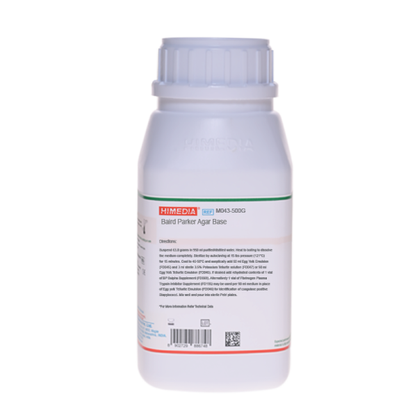
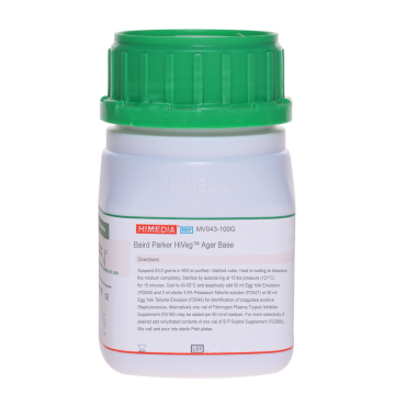
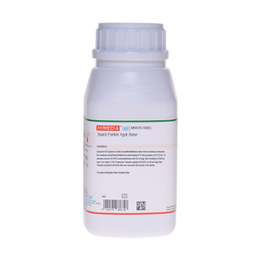
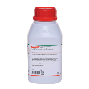
Baird Parker Agar Base
Staphylococci#CC293D
Intended Use
Recommended for the isolation and enumeration of coagulase positive staphylococci from food and clinical samples.
Composition**
| Ingredients | g/L |
|---|---|
| Tryptone | 10.000 |
| HM Peptone B# | 5.000 |
| Yeast extract | 1.000 |
| Glycine | 12.000 |
| Sodium puruvate | 10.000 |
| Lithium chloride | 5.000 |
| Agar | 20.000 |
Final pH (at 25°C): 7.0±0.2
**Formula adjusted, standardized to suit performance parameters # - Equivalent to Beef extract
Directions
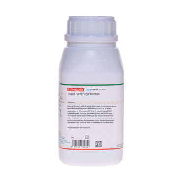
Suspend 63.0 grams in 950 ml purified/ distilled water. Heat to boiling to dissolve the medium completely. Sterilize by autoclaving at 15 lbs pressure (121°C) for 15 minutes. Cool to 50°C and aseptically add 50 ml concentrated Egg Yolk Emulsion (FD045) and 3 ml sterile PTe 3.5% Selective Supplement (1 ml per vial) (FD047) or 50 ml Egg Yolk Tel Emulsion (100 ml per vial) (FD046). For additional selectivity, if desired add rehydrated contents of 1 vial of BP S Selective Supplement (FD069). Alternatively 1 vial of FPT Inhibitor (FD195) may be used per 90 ml medium in place of Egg Yolk Tel Emulsion (100 ml per vial) (FD046) for identification of coagulase, positive Stapylococci. Mix well and pour into sterile Petri plates.
Principle And Interpretation
Baird Parker Agar was developed by Baird Parker (1,2) from the Tellurite-glycine formulation of Zebovitz et al (3) for isolation and enumeration of Staphylococci in food and other material since it allows a good differentiation of coagulase positive strains. A high correlation has been found between the coagulase test and the presence of clear zone of lipolysis in this medium, which is due to the lecithinase of Staphylococci that breakdown, the egg yolk. On the other hand, studies show that almost 100% of coagulase positive Staphylococci are capable of reducing tellurite, which produces black colonies, whereas other Staphylococci cannot always do so. The medium was found to be less inhibitory to Staphylococcus aureus than other media at the same time being more selective (4,5,6). Subsequently the use of Baird-Parker Agar was officially adopted by AOAC International (7) and is recommended in the USP for use in the performance of Microbial Limit Tests (8). Recently, ISO committee has also recommended this medium for the isolation and enumeration of Staphylococci (9).
The identity of Staphylococcus aureus isolated on Baird-Parker Agar must be confirmed with a coagulase reaction. Baird-Parker Agar can also be used to detect coagulase activity by adding fibrinogen plasma (10). FPT Inhibitor (FD195) dissolved in 10 ml sterile distilled water added to 90 ml sterile molten media kept at 45-50°C. On this medium coagulase positive colonies appear white to grey-black surrounded by an opaque zone due to coagulase activity within 24-48 hours incubation at 35°C. Reduction in tellurite is necessary because of absence of egg yolk emulsion. This results in translucent agar and white to grey coloured colonies of Staphylococci. For quantitative results select 20-200 colonies. Count Staphylococcus aureus like colonies and test them for coagulase reaction. Report Staphylococcus aureus per gram of food. Smith and Baird-Parker (11) found that the addition of 50 mg/l Sulphamethazine in the medium, suppresses the growth and swarming of Proteus species.
Tryptone, HM peptone B and yeast extract are sources of nitrogen, carbon, sulphur and vitamins. Sodium pyruvate not only protects injured cells and helps recovery but also stimulates Staphylococcus aureus growth without destroying selectivity. Lithium chloride and potassium tellurite inhibit most of the contaminating microflora except Staphylococcus aureus. The tellurite additive is toxic to egg yolk-clearing strains other than S.aureus and imparts a black colour to the colonies.
Glycine, pyruvate enhances growth of Staphylococcus. With the addition of egg yolk, the medium becomes yellow, opaque. The egg yolk additive, in addition to provide enrichment, aids in the identification process by demonstrating lecithinase activity (egg yolk reaction). A clear zone and grey-black colonies on this medium are diagnostic for coagulase positive Staphylococci. Upon further incubation, an opaque zone is developed around colonies, which can be due to lipolytic activity. When testing the medium, inoculate the material to be examined (0.1 ml per plate of diameter 90-100 mm), incubate at 37°C and take the first reading after 24-26 hours. The colonies of Staphylococcus aureus are black and shiny, with a fine white rim, surrounded by a clear zone. Incubate at 37°C for another 24 hours and perform the coagulase test on the colonies with the above characteristics, which have developed during the further incubation period. Plates should be used on the same day of preparation or within 48 hours, to avoid the loss of definition in the precipitated zones. The basal medium, without the egg yolk or the tellurite, is perfectly stable. Colonies of some contaminating organisms may digest the coagulase halo reaction. Other bacteria may grow on this media but biochemical test will differentiate coagulase positive Staphylococci from the other organisms.
Type of specimen
Clinical samples : Pus, wounds, Food and dairy samples
Specimen Collection and Handling
For food and dairy samples, follow appropriate techniques for sample collection and processing as per guidelines (12,13,14). For clinical samples follow appropriate techniques for handling specimens as per established guidelines (15,16). After use, contaminated materials must be sterilized by autoclaving before discarding.
Warning and Precautions
In Vitro diagnostic use. For professional use only. Read the label before opening the container. Wear protective gloves/protective clothing/eye protection/face protection. Follow good microbiological lab practices while handling specimens and culture. Standard precautions as per established guidelines should be followed while handling clinical specimens. Safety guidelines may be referred in individual safety data sheets.
Limitations
- Though the medium is recommended for detection of coagulase positive Staphylococcus aureus, other bacteria may grow.
- Individual organisms differ in their growth requirement and may show variable growth patterns on the medium.
- Each lot of the medium has been tested with the standard strains, slight variation in growth may be observed depending on the source from where the organism has been isolated.
Performance and Evaluation
Performance of the medium is expected when used as per the direction on the label within the expiry period when stored at recommended temperature.
Quality Control
Appearance Cream to yellow homogeneous free flowing powder
Gelling Firm, comparable with 2.0% agar gel.
Colour and Clarity of prepared medium Basal medium: Yellow coloured clear to slightly opalescent gel. After addition of Egg Yolk Emulsion and Tellurite Emulsion: Yellow coloured opaque gel forms in Petri plates.
Reaction Reaction of 6.3% w/v aqueous solution at 25°C. pH : 7.0±0.2
pH 6.80-7.20
Cultural Response
Cultural response was observed after an incubation at 35-37°C for 24-48 hours. Recovery rate is considered as 100% for bacteria growth on Soyabean Casein Digest Agar.
| Organism | Inoculum (CFU) | Growth | Recovery | Colour of colony | Lecithinase |
|---|---|---|---|---|---|
| Staphylococcus aureus subsp. aureus ATCC 6538 (00032*) | 50-100 | luxuriant | >=50% | grey-black shiny | Positive, opaque zone around the colony |
| Staphylococcus aureus subsp. aureus ATCC 25923 (00034*) | 50-100 | luxuriant | >=50% | grey-black shiny | Positive, opaque zone around the colony |
| Proteus mirabilis ATCC 25933 | 50-100 | good - luxuriant | >=50% | brown - black | Negative |
| Micrococcus luteus ATCC 10240 | 50-100 | poor - good | 30-40% | shades of brown-black (very small) | Negative |
| Staphylococcus epidermidis ATCC 12228 (00036*) | 50-100 | fair-good | 30-40% | black | Negative |
| ** Bacillus spizizenii ATCC 6633 (00003*) | 50-100 | none - poor | 0-10% | dark brown matt | Negative |
| Escherichia coli ATCC 8739 (00012*) | 50-100 | none- poor | 0-10% | large brown black | Negative |
| Escherichia coli ATCC 25922 (00013*) | 50-100 | none- poor | 0-10% | large brown black | Negative |
Key: *Corresponding WDCM numbers, **Formerly known as Bacillus subtilis subsp. spizizenii'
Storage and Shelf Life
Store between 10-30°C in a tightly closed container and the prepared medium at 2 - 8°C. Use before expiry date on the label. On opening, product should be properly stored dry, after tightly capping the bottle in order to prevent lump formation due to the hygroscopic nature of the product. Improper storage of the product may lead to lump formation. Store in dry ventilated area protected from extremes of temperature and sources of ignition Seal the container tightly after use. Product performance is best if used within stated expiry period.
Disposal
User must ensure safe disposal by autoclaving and/or incineration of used or unusable preparations of this product. Follow established laboratory procedures in disposing of infectious materials and material that comes into contact with clinical sample must be decontaminated and disposed of in accordance with current laboratory techniques (15,16).
Reference
- Baird-Parker A. C., 1962, J. Appl. Bacteriol., 25:12.
- Baird-Parker A. C. and Davenport E., 1965, J. Appl. Bacteriol., 28:390.
- Zebovitz E., Evans J. B. and Niven C.F., 1955, J. Bacteriol., 70:686.
- Assoc. off. Anal. Chem., 1971, 54:401.
- Baer, 1971, J. Assoc. Off. Anal. Chem., 54:732.
- Tardio and Baer, 1971, J. Assoc. Off. Anal. Chem., 54:728.
- Horwitz (Ed.), 2000, Official methods of analysis of AOAC International, 17th Ed., Vol. I., AOAC International, Gaithersburg, MD.
- The United States Pharmacopoeia-National Formulatory (USP-NF), 2022.
- International Organization for Standardization (ISO), 1983, Draft ISO/DIS 6888.
- Beckers N. J. et al, 1984, Can. J. Microbiol., 30:470.
- Smith B. A. and Baird-Parker A.C., 1964, J. Appl. Bacteriol., 27:78.
- Salfinger Y., and Tortorello M.L., 2015, Compendium of Methods for the Microbiological Examination of Foods, 5th Ed., American Public Health Association, Washington, D.C.
- Wehr H. M. and Frank J. H., 2004, Standard Methods for the Microbiological Examination of Dairy Products, 17th Ed., APHA Inc., Washington, D.C.
- American Public Health Association, Standard Methods for the Examination of Dairy Products, 1978, 14th Ed., Washington.
- Isenberg, H.D. Clinical Microbiology Procedures Handbook 2nd Edition.
- Jorgensen, J.H., Pfaller, M.A., Carroll, K.C., Funke, G., Landry, M.L., Richter, S.S and Warnock., D.W. (2015) Manual of Clinical Microbiology, 11th Edition. Vol. 1.
| Product Name | Baird Parker Agar Base |
|---|---|
| SKU | M043 |
| Product Type | Regular |
| Physical Form | Powder |
| Origin | Animal |
| Packaging type | HDPE |
| References | 1. Baird-Parker A. C., 1962, J. Appl. Bacteriol., 25:12. 2.Baird-Parker A. C. and Davenport E., 1965, J. Appl. Bacteriol., 28:390. 3.Zebovitz E., Evans J. B. and Niven C.F., 1955, J. Bacteriol., 70:686 . 4.Tardio and Baer, 1971, J. Assoc. Off. Anal. Chem., 54:728. 5.Baer, 1971, J. Assoc. Off. Anal. Chem., 54:732. 6.Assoc. off. Anal. Chem., 1971, 54:401. 7.Horwitz (Ed.), 2000, Official methods of analysis of AOAC International, 17th Ed., Vol. I., AOAC International,Gaithersburg, MD. 8.The United States Pharmacopoeia, 2018, The United States Pharmacopoeial Convention. Rockville, MD. 9.International Organization for Standardization (ISO), 1983, Draft ISO/DIS 6888. 10.Smith B. A. and Baird-Parker A.C., 1964, J. Appl. Bacteriol., 27:78. 11. Beckers N. J. et al, 1984, Can. J. Microbiol., 30:470.1 2.American Public Health Association, Standard Methods for the Examination of Dairy Products, 1978, 14th Ed., WashingtonD.C. 13.Salfinger Y., and Tortorello M.L. Fifth (Ed.), 2015, Compendium of Methods for the Microbiological Examination ofFoods, 5th Ed., American Public Health Association, Washington, D.C. 14..Wehr H. M. and Frank J. H., 2004, Standard Methods for the Microbiological Examination of Dairy Products, 17th Ed.,APHA Inc., Washington, D.C. 15.Isenberg, H.D. Clinical Microbiology Procedures Handb0ook. 2nd Edition. 16.Jorgensen,J.H., Pfaller , M.A., Carroll, K.C., Funke, G., Landry, M.L., Richter, S.S and Warnock., D.W. (2015)Manual of Clinical Microbiology, 11th Edition. Vol. 1. |
| Customized Product Available | No |


