 Your enquiry has been submitted
Something problem with the server. Try again.
Recommended for primary isolation and identification of Candida species.
| Ingredients | g / L |
|---|---|
| Peptone | 10.000 |
| Yeast extract | 1.000 |
| Dextrose (Glucose) | 40.000 |
| Bromocresol green | 0.020 |
| Agar | 15.000 |
Final pH ( at 25°C): 6.1±0.2
**Formula adjusted, standardized to suit performance parameters
Suspend 66.02 grams in 1000 ml purified / distilled water. Heat to boiling to dissolve the medium completely. Sterilize by autoclaving at 15 lbs pressure (121°C) for 15 minutes. Cool to 45-50°C and add sterile neomycin to a concentration of 500 µg/ml of medium. Mix well before pouring into sterile Petri plates.
Candida albicans is most frequently isolated from clinical specimens. Species of Candida, other than C. albicans are normal flora of cutaneous and mucocutaneous surfaces and are only rarely incriminated as agents of clinical disease (1). Of the many media used for isolating and differentiating Candida, Pagano Levin Base (M1390) employes TTC (Triphenyl Tetrazolium Chloride) as an indicator. Harold and Snyder (2) observed that the TTC used greatly retards the growth of some Candida species, while completely inhibiting the rest. Therefore to overcome this difficulty, they formulated Candida BCG Agar, which employs bromocresol green instead of TTC as the indicator.
Candida BCG Agar Base is used to obtain pure yeast colonies from mixed cultures on the basis of colony morphology (3, 4). Peptone along with yeast extract and dextrose serve as sources of essential nutrients, amino acids and vitamins. Dextrose also serves as a source of energy by being the fermentable carbohydrate. Bromocresol green is non-toxic indicator incorporated to visualize the fermentation reaction. Selectivity is obtained by the addition of neomycin. Neomycin is incorporated to inhibit gram-negative bacteria and some gram-positive bacteria. Neomycin is an aminoglycoside antibiotic that is active against aerobic and facultatively anaerobic gram-negative bacteria and certain gram-positive bacteria. Bromocresol green is the indicator. Acid production due to fermentation lowers the pH of the medium and subsequently the colour of medium changes to yellow, indicated by yellow zones around the dextrose-fermenting colonies. C.albicans appears as blunt conical colonies with smooth edges and yellow to blue green colour. Other Candida species appear as smooth to rough colonies, with either convex or cone shaped colonies (5). Standard methods should be followed for inoculating the plates of Candida BCG Agar.
Presumptive Candida colonies should be further identified by gram staining, biochemical and serological testing (6,7,8).
Clinical samples - skin scraping from the infected body site.
For clinical samples follow appropriate techniques for handling specimens as per established guidelines (9,10). After use, contaminated materials must be sterilized by autoclaving before discarding.
In Vitro diagnostic Use only. For professional use only. Read the label before opening the container. Wear protective gloves/protective clothing/eye protection/ face protection. Follow good microbiological lab practices while handling specimens and culture. Standard precautions as per established guidelines should be followed while handling clinical specimens. Safety guidelines may be referred in individual safety data sheets.
Performance of the medium is expected when used as per the direction on the label within the expiry period when stored at recommended temperature.
Appearance: Cream to light green homogeneous free flowing powder
Gelling: Firm, comparable with 1.5% Agar gel
Colour and Clarity of prepared medium: Bluish green coloured, clear to slightly opalescent gel forms in Petri plates
Reaction: Reaction of 6.6% w/v aqueous solution at 25°C. pH : 6.1±0.2
pH: 5.90-6.30
Cultural Response: Cultural characteristics observed with added sterile Neomycin (500 mcg/ml of medium) after an incubation at 25-30°C for 24-48 hours.
| Organism | Inoculum (CFU) | Growth | Recovery | Colour of medium |
|---|---|---|---|---|
| Candida albicans ATCC 10231 (00054*) | 50-100 | good-luxuriant | >=50% | yellow |
| Candida glabrata ATCC 15126 | 50-100 | good-luxuriant | >=50% | yellow |
| Candida kruisei ATCC 24408 | 50-100 | good-luxuriant | >=50% | yellow |
| Candida tropicalis ATCC 1369 | 50-100 | good-luxuriant | >50% | yellow |
| Escherichia coli ATCC 25922 (00013*) | >=104 | inhibited | 0% | |
| Staphylococcus aureus subsp. aureus ATCC 25923 (00034*) | >=104 | inhibited | 0% |
Key : *Corresponding WDCM numbers.
Store between 10-30°C in a tightly closed container and the prepared medium at 20-30°C. Use before expiry date on the label. On opening, product should be properly stored dry, after tightly capping the bottle in order to prevent lump formation due to the hygroscopic nature of the product. Improper storage of the product may lead to lump formation. Store in dry ventilated area protected from extremes of temperature and sources of ignition. Seal the container tightly after use. Product performance is best if used within stated expiry period.
User must ensure safe disposal by autoclaving and/or incineration of used or unusable preparations of this product. Follow established laboratory procedures in disposing of infectious materials and material that comes into contact with clinical sample must be decontaminated and disposed of in accordance with current laboratory techniques (9,10).
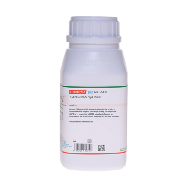
| Product Name | Candida BCG Agar Base |
|---|---|
| SKU | M355 |
| Product Type | Regular |
| Physical Form | Powder |
| Origin | Animal |
| Packaging type | HDPE |
| References | 1.Association of Analytical Chemists (AOAC), 1960, 9th ed., Published by AOAC International. 2.Journal of AOAC, 1964, 47:176. 3.Methods of Analysis AOAC, 1980, 13th ed. |
| Customized Product Available | No |