 Your enquiry has been submitted
Something problem with the server. Try again.
Your enquiry has been submitted
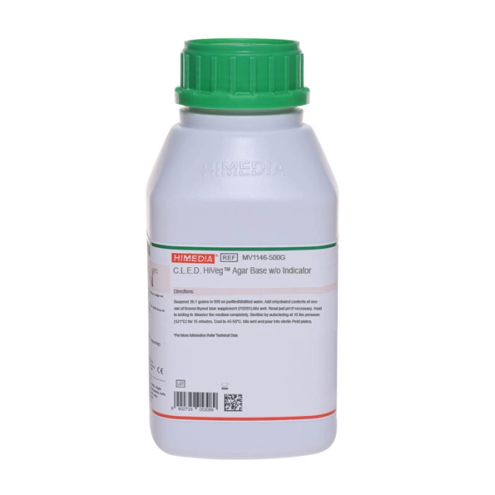
Recommended for isolation and differentiation of urinary pathogens on the basis of lactose fermentation.
| Ingredients | g / L |
|---|---|
| Peptone | 4.000 |
| HM peptone B # | 3.000 |
| Tryptone | 4.000 |
| Lactose | 10.000 |
| L-Cystine | 0.128 |
| Bromothymol blue | 0.020 |
| Andrade indicator | 0.100 |
| Agar | 15.000 |
Final pH (at 25°C): 7.5±0.2
**Formula adjusted, standardized to suit performance parameters
# - Equivalent to Beef extract
Suspend 36.25 grams in 1000 ml purified/distilled water. Heat to boiling to dissolve the medium completely. Sterilize by autoclaving at 15 lbs pressure (121°C) for 15 minutes. Cool to 45-50°C. Mix well and pour into sterile Petri plates.
Sandys reported a new technique where the swarming of Proteus on an agar medium could be prevented by restricting the electrolyte content in the culture medium (1). Sandys Medium was modified by Mackey and Sandys (2), by replacing mannitol with lactose and sucrose and elevating the concentration of agar and bromothymol blue. The same authors further modified this medium by retaining the lactose (deleting sucrose) and by including L-cystine for promoting the growth of cystine-dependent dwarf coliform colony (3). This later modified medium was designated as C.L.E.D. (Cystine- Lactose Electrolyte-Deficient) Medium. This medium is recommended for use in urinary bacteriology, promoting the growth of all urinary pathogens. C.L.E.D. Medium is also recommended for dip stick procedures and as dip inoculum transport medium for urine specimens (2,3,4).
C.L.E.D. Medium was further modified by Bevis (5) by incorporation of Andrades indicator. This medium provides sharper differentiation between lactose-fermenters (LF) and lactose-non-fermenters (NLF) (5). Addition of Andrades indicator enhances the appearance of colony and aids in the identification of microorganisms.
At different pH values, the colour of the medium varies from the standard medium, which is well documented by Bevis (5).
| pH | Colour of C.L.E.D. medium |
|---|---|
| 7.4 | deep blue |
| 7.0 | bluish grey |
| 6.8 | pale grey |
| 6.6 | pinkish grey |
| 6.4 | bright red with whitish tinge |
| 6.0 | bright red |
For better results, the medium should not be incubated for more than 24 hours because if lactose fermenters predominate, the entire medium may turn pink masking the presence of non-lactose fermenters. Inoculate the medium immediately after urine collection. Shigella species may not grow on this medium. Prior initiation of antibiotic therapy, low urine pH (less than 5) etc. may result in low urine count from infected patients.
Clinical: Urine sample
For clinical samples follow appropriate techniques for handling specimens as per established guidelines (6,7). After use, contaminated materials must be sterilized by autoclaving before discarding.
In Vitro diagnostic use only. For professional use only. Read the label before opening the container. Wear protective gloves/protective clothing/eye protection/face protection. Follow good microbiological lab practices while handling specimens and culture. Standard precautions as per established guidelines should be followed while handling clinical specimens. Safety guidelines may be referred in individual safety data sheets.
Performance of the medium is expected when used as per the direction on the label within the expiry period when stored at recommended temperature.
Appearance Light yellow to grayish yellow homogeneous free flowing powder
Gelling Firm, comparable with 1.5% Agar gel
Colour and Clarity of prepared medium Greenish blue clear to slightly opalescent gel forms in Petri plates
Reaction Reaction of 3.62% w/v aqueous solution at 25°C. pH : 7.5±0.2
pH 7.30-7.70
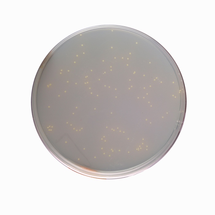
Cultural Response Cultural characteristics observed after an incubation at 35-37°C for 18-24 hours
| Organism | Inoculum (CFU) | Growth | Recovery | Colour of colony |
|---|---|---|---|---|
| # Klebsiella aerogenes ATCC 13048 (00175*) | 50-100 | good-luxuriant | >=70% | greyish green |
| Escherichia coli ATCC 25922 (00013*) | 50-100 | good-luxuriant | >=70% | bright pink with pink halo |
| Enterococcus faecalis ATCC 29212 (00087*) | 50-100 | good-luxuriant | >=70% | orange-yellow or greenish |
| Proteus mirabilis ATCC 25933 | 50-100 | good-luxuriant | >=70% | blue-green |
| Staphylococcus aureus subsp. aureus ATCC 25923 (00034*) | 50-100 | good-luxuriant | >=70% | golden-yellow |
| Streptococcus pyogenes ATCC 19615 | 50-100 | good-luxuriant | >=70% | greyish green |
Key : *Corresponding WDCM numbers. # - Formerly known as Enterobacter aerogenes
Store between 10-30°C in a tightly closed container and the prepared medium at 20-30°C. Use before expiry date on the label. On opening, product should be properly stored dry, after tightly capping the bottle in order to prevent lump formation due to the hygroscopic nature of the product. Improper storage of the product may lead to lump formation. Store in dry ventilated area protected from extremes of temperature and sources of ignition Seal the container tightly after use. Product performance is best if used within stated expiry period.
User must ensure safe disposal by autoclaving and/or incineration of used or unusable preparations of this product. Follow established laboratory procedures in disposing of infectious materials and material that comes into contact with clinical sample must be decontaminated and disposed of in accordance with current laboratory techniques (6,7).
| Product Name | C.L.E.D.Agar w/ Andrade Indicator |
|---|---|
| SKU | M352 |
| Product Type | Regular |
| Physical Form | Powder |
| Origin | Animal |
| Packaging type | HDPE |
| References | 1. Laboratory Methods in Anaerobic Bacteriology, 1974, CDC Laboratory Manual, U.S. Dept. HEW, Pub. No. 74-8262. 2.Atlas, R. M., 2004, A Handbook of Microbiological Media, 3rd Ed, CRC Press. 3.MacFaddin, J. F., 1985, (Ed), Media for Isolation-Cultivation-Identification of Medical Bacteria. Vol. I., Williams and Wilkins, Baltimore. 4.Dowell V. R. Jr., Lombad G. L., Thompson F. S., Armfield A. Y., Media for Isolation, Characterization and Identification of Obligately Anaerobic Bacteria, USDHEW Atlanta, CA: Centers for Disease Control, 1977:22 5.Washington J. A., Laboratory Procedures in Clinical Microbiology, Cd 2 New York: Springer-Verlag, 1985: 774, 801-802. |
| Customized Product Available | No |