 Your enquiry has been submitted
Something problem with the server. Try again.
Your enquiry has been submitted
Recommended for primary isolation and identification of pathogenic Staphylococci from clinical specimens or for classifying pure cultures.
| Ingredients | g / L |
|---|---|
| HI infusion$ | 5.000 |
| Tryptone | 10.500 |
| Soya peptone | 3.500 |
| Sodium chloride | 3.500 |
| Mannitol | 10.000 |
| Bromo cresol purple | 0.020 |
| Agar | 14.500 |
Final pH (at 25°C): 7.4±0.2
**Formula adjusted, standardized to suit performance parameters
$ - Equivalent to Beef heart infusion (solids)
Suspend 47.02 grams in 1000 ml purified/distilled water. Heat to boiling to dissolve the medium completely. Sterilize by autoclaving ∆ 118°C-121°C for 15 minutes. Cool to 45-50°C. Add 7-15% v/v sterile pretested, rabbit plasma to the basal medium. Mix well and pour into sterile Petri plates.
∆ corresponds to 12-15 lbs pressure.
The genus Staphylococcus comprises 28 accepted or proposed species, 14 of which may be encountered in human clinical specimens. Staphylococci are generally found on the skin and mucous membranes of humans and other animals. Some of the pathogenic staphylococci in both humans and animals produce an enzyme called coagulase and detection of this enzyme is used in the laboratory to identify these organisms (1).
These media are used for the isolation of Staphylococcus aureus from clinical specimens and for differentiation of S.aureus from other species on the basis of coagulase production and mannitol fermentation. Chapman for the first time introduced a medium for selective isolation and differentiation of Staphylococci (1). Tellurite-glycine media were designed by Zebovitz et al (2) and Marwin (3) for selectively isolating coagulase-positive Staphylococcal species. Present medium is based on Esber and Faulconer formulation (4). Mutant or old cultures of S.aureus may be weak coagulase producers. They should be freshly sub cultured and rechecked. Escherichia coli ferments mannitol and may be weakly coagulase positive. Coagulase production is dependent on the presence of a fermentable sugar like mannitol in this case. It is also dependent on the presence of a protein factor in the HI infusion and blood plasma (5). When mannitol is fermented, the pH of the medium surrounding the coagulase positive colonies drops. This drop in pH is indicated by the change in colour of the bromocresol purple indicator, which turns yellow and exhibits yellow zones around the colonies.
An opaque area of coagulated plasma forms around the colonies of coagulase positive organisms. Staphylococcus epidermidis is coagulase negative and mannitol non-fermenting species, which does not change the colour of the medium. Coagulase negative species may ferment mannitol and produce a yellow zone around the colonies but an opaque zone will not be formed.
Clinical samples :skin and mucous membranes
For clinical samples follow appropriate techniques for handling specimens as per established guidelines (2,3). After use, contaminated materials must be sterilized by autoclaving before discarding.
In Vitro diagnostic use only. For professional use only. Read the label before opening the container. Wear protective gloves/protective clothing/eye protection/face protection. Follow good microbiological lab practices while handling specimens and culture. Standard precautions as per established guidelines should be followed while handling clinical specimens. Safety guidelines may be referred in individual safety data sheets.
Performance of the medium is expected when used as per the direction on the label within the expiry period when stored at recommended temperature.
Appearance: Light yellow to light grey homogeneous free flowing powder
Gelling: Firm, comparable with 1.45% Agar gel
Colour and Clarity of prepared medium: Purple coloured, slightly opalescent gel forms in Petri plates
Reaction: Reaction of 4.7% w/v aqueous solution at 25°C. pH : 7.4±0.2
pH: 7.20-7.60
Cultural Response: Cultural characteristics observed with added 7-15% v/v sterile pretested, rabbit plasma at 35-37°C for 18-48 hours.
| Organism | Inoculum (CFU) | Growth | Recovery | Mannitol fermentation | Coagulase production |
|---|---|---|---|---|---|
| Staphylococcus epidermidis ATCC 12228 (00036*) | 50-100 | luxuriant | >=70% | negative reaction, purple colour | negative reaction, no opaque zone formation |
| Staphylococcus aureus subsp. aureus ATCC 25923 (00034*) | 50-100 | luxuriant | >=70% | positive reaction, yellow colour | positive reaction, colonies surrounded by opaque zone |
Key : *Corresponding WDCM numbers.
Store between 10-30°C in a tightly closed container and the prepared medium at 2-8°C. Use before expiry date on the label. On opening, product should be properly stored dry, after tightly capping the bottle in order to prevent lump formation due to the hygroscopic nature of the product. Improper storage of the product may lead to lump formation. Store in dry ventilated area protected from extremes of temperature and sources of ignition Seal the container tightly after use. Use before expiry date on the label. Product performance is best if used within stated expiry period.
User must ensure safe disposal by autoclaving and/or incineration of used or unusable preparations of this product. Follow established laboratory procedures in disposing of infectious materials and material that comes into contact with clinical sample must be decontaminated and disposed of in accordance with current laboratory techniques (2,3).
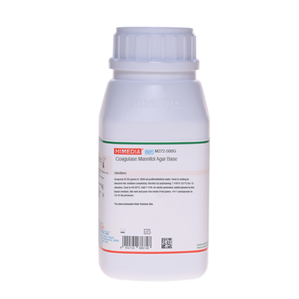
| Product Name | Coagulase Mannitol Agar Base |
|---|---|
| SKU | M272 |
| Product Type | Regular |
| Physical Form | Powder |
| Origin | Animal |
| Packaging type | HDPE |
| References | 1. MacFaddin J., 1985, Media for Isolation-Cultivation-Identification-Maintenance of Medical Bacteria, Vol. I, Williams andWilkins, Baltimore. 2.Finegold and Baron, 1986, Bailey and Scotts Diagnostic Microbiology, 7th ed., The C.V. Mosby Co., St. Louis. 3.Ewing, 1986, Edwards and Ewings Identification of Enterobacteriaceae, 4th ed., Elsevier Science Publishing Co., Inc., NewYork. |
| Customized Product Available | No |